Download presentation
Presentation is loading. Please wait.

1
Prof. Liu Jianhua PhD. MS. DDS. Dept. of Oral-Maxillofacial Surg. & Dentistry First affiliated hospital Zhejiang university school of medicine

2
Tumors in oral & maxillo-facial region Naming : original tissue, biological type, position. Such as: squamous cell carcinoma of right tongue Critical t. : biologically benign tumor, but has malignant tendency and infiltrating growth. such as : ameloblastoma, mixed tumor, papiloma.

3
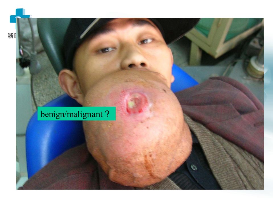
A mass in left submandibular region for 5 years, slow growing, with no symptom. Diagnosis? Further examination?

4
Left tongue ulcer 3 months, painful, Poor condition of oral cavity, what to do next?

5
Black lesion in soft palate, grows rapidly recent 3 months, shall we make a biopsy?

6
manifestations & diagnosis Judgement of a mass on patient : 1 ) tumor / not tumor(inflam. TB, malformation, etc.) 2 ) benign / malignant
 2 ) benign / malignant.")
7
Differentiation benign tumor malignant tumor Age any age carcinoma/agedness ; sarcoma/young Growth speed slow faster Growth manner bulge invasive Boundary smooth , movable unclear , unmovable Symptom generally no painful/numbness/funct.limmit Metastasis no maybe Body affect generally no organ destroy/system destroy Histology struct. well differentiated worse diff./abn. nuclear close to norm.cell/tissue division ;

8
benign/malignant ?

10
Means of diagnosis 1 ) case history : arise , progress 2 ) clinical examine : look 、 touch 、 listen to(with stethoscope ) 3 ) image : X-ray , panoramic radiograph , CT , MRI , DSA , B- ultrasonic , PET/CT 4 ) puncture : for liquid 5 ) biopsy : no melanoma/parotid g. 6 ) tumor markers : CEA, AFP, CA50/125/153/242/724 etc.
 tumor markers : CEA, AFP, CA50/125/153/242/724 etc..")
11
treatment 1 ) treatment principle : benign tumor— surgery first ; malignant t.—combined 2 ) methods : Surgery. : safe edge—1 cm for malignacy Radiotherapy. : oral preparation—remove ill tooth and metal crown; fill decayed tooth; tooth cleaning---(to prevent jaw infection, necrosis, redial beam focus) Chemotherapy : before,during, post-operation others : thermotherapy./immunother. /herbs/ biotherapy
 Chemotherapy : before,during, post-operation others : thermotherapy./immunother. /herbs/ biotherapy.")
12
Prevention of oral cancer 1.eliminate or reduce carcinogen : incomplete tooth root or crown 、 displaced tooth 、 sharp tooth edge 、 poor denture ; hot food 、 tobacco and alcohol ; environment contamination ( air pollution , paint , glue ); psycho- problem. 2.treat precancerous lesion in time : mucosa erythema 、 leukoplakia 、 oral lichen planus 、 papilloma etc. 3.investigate vulnerable population and give education

13
question : how to prevent oral cancers ?

14
第二节 Oral & Maxillofacial cyst 口腔颌面部囊肿

15
Commonness of Cysts 囊肿共性 有囊壁 wall 、囊内容物 content (液 体 / 固形物 liquid/solid ) 边界清楚 缓慢增大 颌骨囊肿可造成骨组织压迫性吸收 expansile absorbing
 边界清楚 缓慢增大 颌骨囊肿可造成骨组织压迫性吸收 expansile absorbing")
16
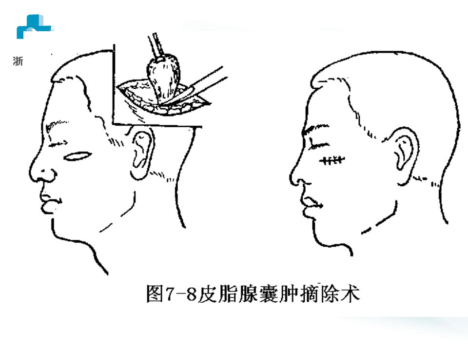
软组织囊肿 一、皮脂腺囊肿 sebaceous cyst 病因 pathogeny :皮脂腺 sebaceous gland 排泄管 阻塞,分泌物蓄积 ---- 潴留性 retentive 囊肿。 临床表现:与皮肤粘连。色素点 pigment dot 。 恶性变 ---- 皮脂腺癌。 治疗:梭形 shuttle 切口 -- 疤痕。二次法(刘氏)
")
18
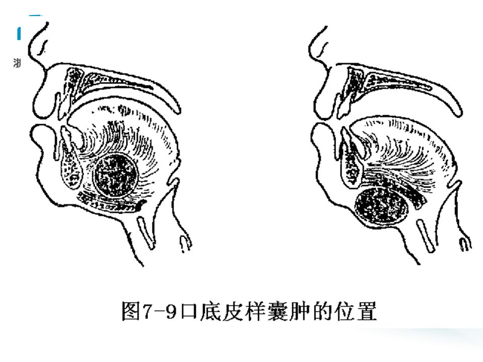
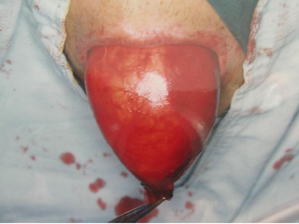
二、皮样、表皮样囊肿 dermoid / epidermoid cyst 病因: 胚胎 embryo 期上皮残留 remnant 、 后天植入(手术、外伤) 临床表现:青少年。皮样 ---- 口底、颏下。 表皮样 ---- 面部。与皮肤无粘连。面团样 - --- 上皮细胞、毛囊、毛发、皮脂腺。 治疗: 手术摘除。下颌舌骨肌、颏舌骨 肌 ---- 上、下进路不同。
 临床表现:青少年。皮样 ---- 口底、颏下。 表皮样 ---- 面部。与皮肤无粘连。面团样 上皮细胞、毛囊、毛发、皮脂腺。 治疗: 手术摘除。下颌舌骨肌、颏舌骨 肌 ---- 上、下进路不同。")
22
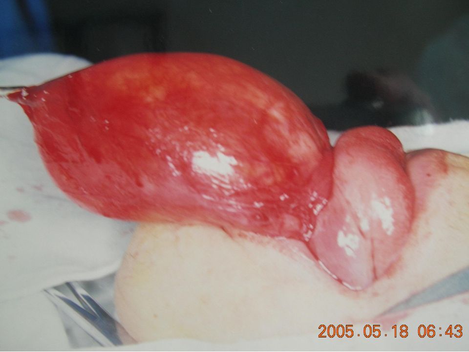
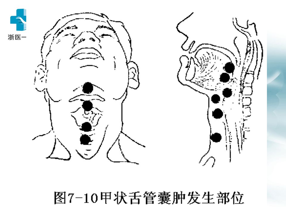
三、甲状舌管囊肿 thyroglossal tract cyst 病因:胚胎第四周 ---- 甲状腺始基 ---- 甲状 舌管 ---- 第六周:管消失,留下甲状腺、 舌盲孔。管残留 ---- 囊肿。下移障碍 ---- 异 位甲状腺。 临床表现: 1-10 岁 > 成年。 舌盲孔 ---- 胸 骨切迹之间。舌骨上下最多。圆形,质 中,囊性感,随吞咽、伸舌运动,穿刺 -- -- 无色 / 有色液体, B 超。感染 ---- 瘘 ---- 癌 变。

24
(鉴别诊断) 舌异位甲状腺:舌根部,蓝紫色,柔软, “ 含橄榄 ” 音。 1 、迷走甲状腺:除异位外,颈部无甲状腺。 2 、副甲状腺:颈部也有,舌根也有。 * 碘 -131/ 锝 -99 扫描为可靠依据。 治疗 囊肿 + 舌骨中段一起切除。 误切迷走甲状腺 ---- 终身服药。
 舌异位甲状腺:舌根部,蓝紫色,柔软, 含橄榄 音。 1 、迷走甲状腺:除异位外,颈部无甲状腺。 2 、副甲状腺:颈部也有,舌根也有。 * 碘 -131/ 锝 -99 扫描为可靠依据。 治疗 囊肿 + 舌骨中段一起切除。 误切迷走甲状腺 ---- 终身服药。")
25
四 鳃裂囊肿 branchial cleft cyst 病因: 胚胎第三周 ----5 对鳃弓 ----4 对鳃裂 ---- 鳃 裂上皮残余 ---- 囊肿 临床表现: 20-50 岁(第一鳃裂年龄较小)。 质软,波动感 fluctuate ,上呼吸道感染 ---- 增大, 恶变 ---- 鳃裂癌,感染 ---- 鳃裂瘘 fistula (有外口 无内口),先天瘘 congenital---- 原发性鳃裂瘘 (有外口也有内口),穿刺 ---- 有色 / 无色;含 / 不含胆固醇液, B 超。
。 质软,波动感 fluctuate ,上呼吸道感染 ---- 增大, 恶变 ---- 鳃裂癌,感染 ---- 鳃裂瘘 fistula (有外口 无内口),先天瘘 congenital---- 原发性鳃裂瘘 (有外口也有内口),穿刺 ---- 有色 / 无色;含 / 不含胆固醇液, B 超。")
26
二、颌骨囊肿 maxillary cyst 牙源性颌骨囊肿 odontogenic jaw cyst 造牙组织 / 牙 ---- 演变而来(根尖、含牙、 角化、始基) 非牙源性颌骨囊肿 non-odontogenic 胚胎期面突残余上皮 ---- 面裂囊肿 创伤 ---- 血外渗性囊肿 intra-bleeding
 非牙源性颌骨囊肿 non-odontogenic 胚胎期面突残余上皮 ---- 面裂囊肿 创伤 ---- 血外渗性囊肿 intra-bleeding")
27
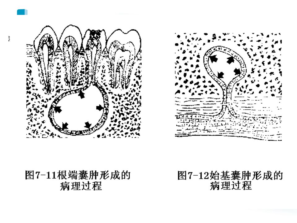
牙源性颌骨囊肿 odontogenic jaw cyst 根尖囊肿( radicular cyst) 慢性根尖周炎 ---- 肉芽肿 ---- 牙周膜上皮残 余 ---- 上皮团块 ---- 液化 残余囊肿( residual cyst) :拔牙后肉芽肿 残留所致
 慢性根尖周炎 ---- 肉芽肿 ---- 牙周膜上皮残 余 ---- 上皮团块 ---- 液化 残余囊肿( residual cyst) :拔牙后肉芽肿 残留所致")
28
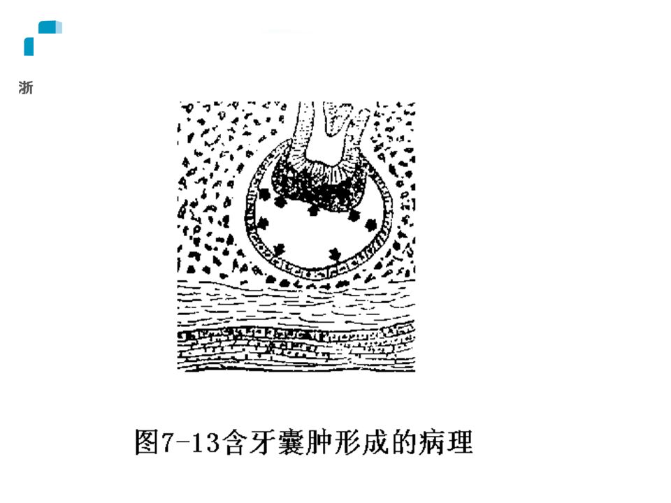
始基囊肿 (primordial cyst) 造釉器发育早期 ---- 星网层变性 ---- 渗出 含牙囊肿 (dentigerous cyst) 牙冠表面与缩余釉上皮之间 ---- 渗出 角化囊肿 (keratocyst) 原始牙胚 / 牙板残余 ---- 增生(含角化物, 壁厚,子囊 / 上皮岛)
 造釉器发育早期 ---- 星网层变性 ---- 渗出 含牙囊肿 (dentigerous cyst) 牙冠表面与缩余釉上皮之间 ---- 渗出 角化囊肿 (keratocyst) 原始牙胚 / 牙板残余 ---- 增生(含角化物, 壁厚,子囊 / 上皮岛)")
31
临床表现 clinical characteristics 缓慢、膨胀性生长 ---- 颌骨坚硬肿块 ---- 乒 乓感 ---- 波动感 ---- 骨折

32
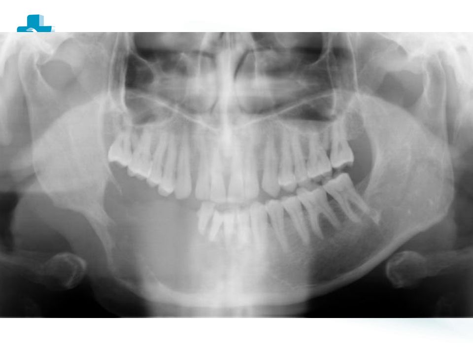
诊断 穿刺 草黄色液体 X 线 根尖囊肿 ---- 根尖在囊内,死髓 始基囊肿 ---- 下 8 区 / 升支部好发 含牙囊肿 ---- 牙冠在囊内 角化囊肿 ---- 下 8 区 / 升支部好发

33
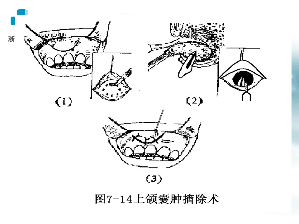
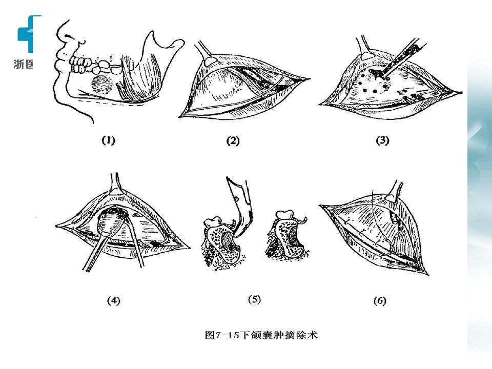
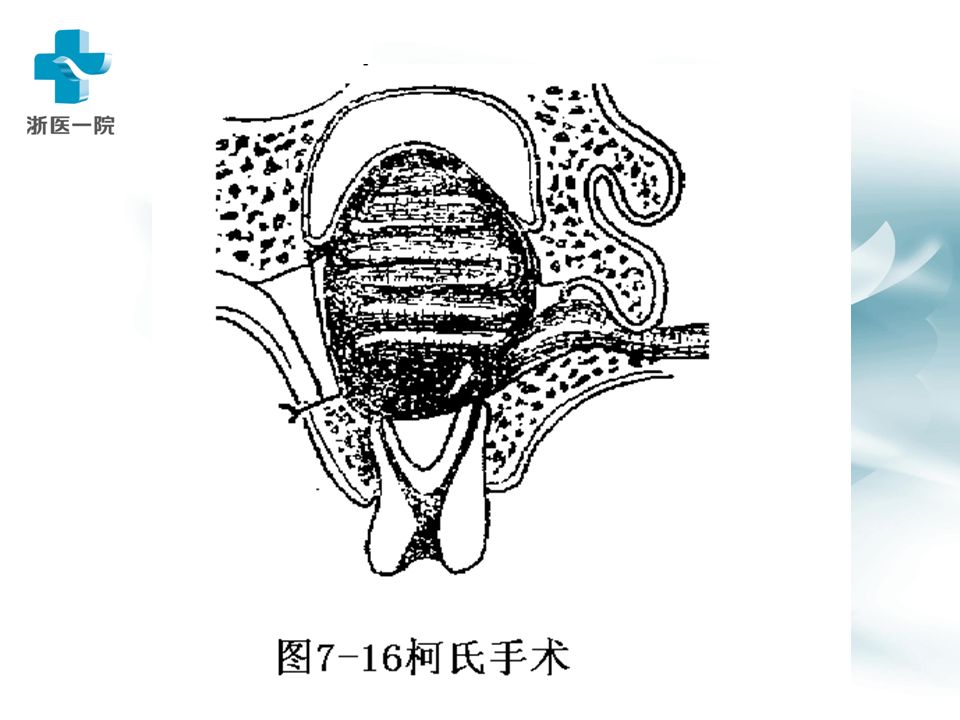
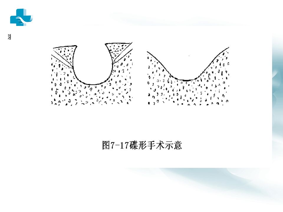
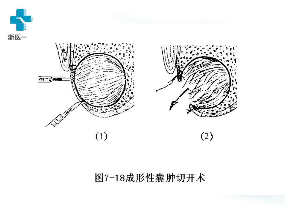
治疗 手术摘除 局麻 / 全麻,口内 / 口外 角化囊肿易复发 / 癌变:石炭酸 / 硝酸银腐 蚀骨壁 死腔消除

39
第三节 良性肿瘤和瘤样病变 benign tumor & tumor-like lesion 口腔颌面部软 / 硬组织内的良性、实质性、 占位性病变,生长缓慢(一般以年为单 位)。
。")
40
一、 瘤样病变

41
色素痣 nevus

42
皮肤构成:图示 色素痣起源:基底层黑色素细胞团。 皮内痣 --- 真皮内,分化高的小痣细胞团, 如雀斑样。 交界痣 --- 表皮真皮交界;大痣细胞;可 恶性变 --- 恶黑。 复合痣 --- 表皮 + 真皮;大痣 + 小痣细胞, 如毛痣。

43
牙龈瘤 (epulis) 病因: 机械 / 慢性炎症刺激 --- 牙龈肿块(非真 性肿瘤) 肉芽肿型 --- 炎症细胞 + 毛细血管为主,易出血 纤维型 --- 纤维组织 + 纤维母细胞,不易出血 血管型 --- 似血管瘤,极易出血(妊娠性龈瘤) 治疗原则: 易复发 --- 去除病变 + 牙 + 邻近骨 组织
 病因: 机械 / 慢性炎症刺激 --- 牙龈肿块(非真 性肿瘤) 肉芽肿型 --- 炎症细胞 + 毛细血管为主,易出血 纤维型 --- 纤维组织 + 纤维母细胞,不易出血 血管型 --- 似血管瘤,极易出血(妊娠性龈瘤) 治疗原则: 易复发 --- 去除病变 + 牙 + 邻近骨 组织")
44
二、良性肿瘤

45
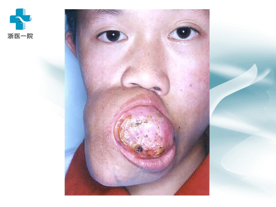
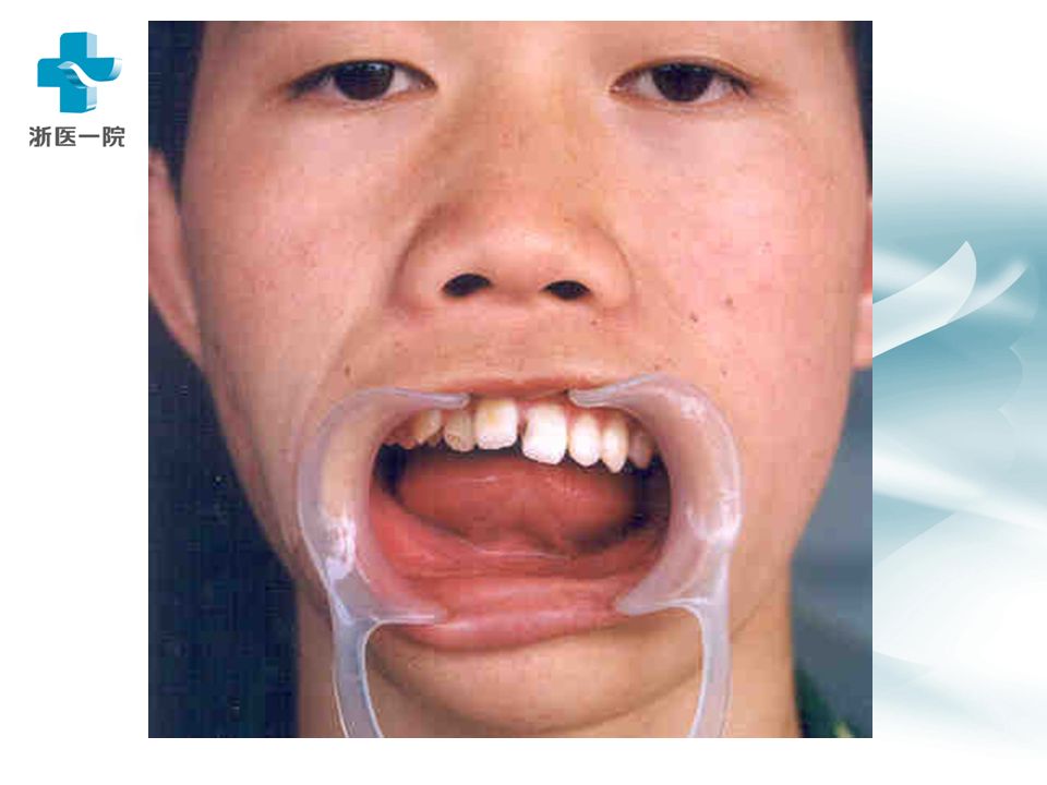
(一)、成釉质细胞瘤 ( ameloblastoma) 多数人认为由造釉器或牙板上皮发生而来。剖面实质性 (多) / 囊性(褐色液),或混合。 临床表现:青壮年多,下颌体 / 角多,缓慢增大,大到 一定程度 ---- 各种临床表现(牙、骨、神经受累)。可 恶变(少)。 诊断: X 片:蜂房状 ---- 多房性囊肿样阴影,边缘不齐, 半月型切迹,根尖吸收。 治疗:易复发,须在边界外 0.5cm 切除。植骨准备。 问题:成牙釉质细胞瘤有那些临床特点?
、成釉质细胞瘤 ( ameloblastoma) 多数人认为由造釉器或牙板上皮发生而来。剖面实质性 (多) / 囊性(褐色液),或混合。 临床表现:青壮年多,下颌体 / 角多,缓慢增大,大到 一定程度 ---- 各种临床表现(牙、骨、神经受累)。可 恶变(少)。 诊断: X 片:蜂房状 ---- 多房性囊肿样阴影,边缘不齐, 半月型切迹,根尖吸收。 治疗:易复发,须在边界外 0.5cm 切除。植骨准备。 问题:成牙釉质细胞瘤有那些临床特点?")
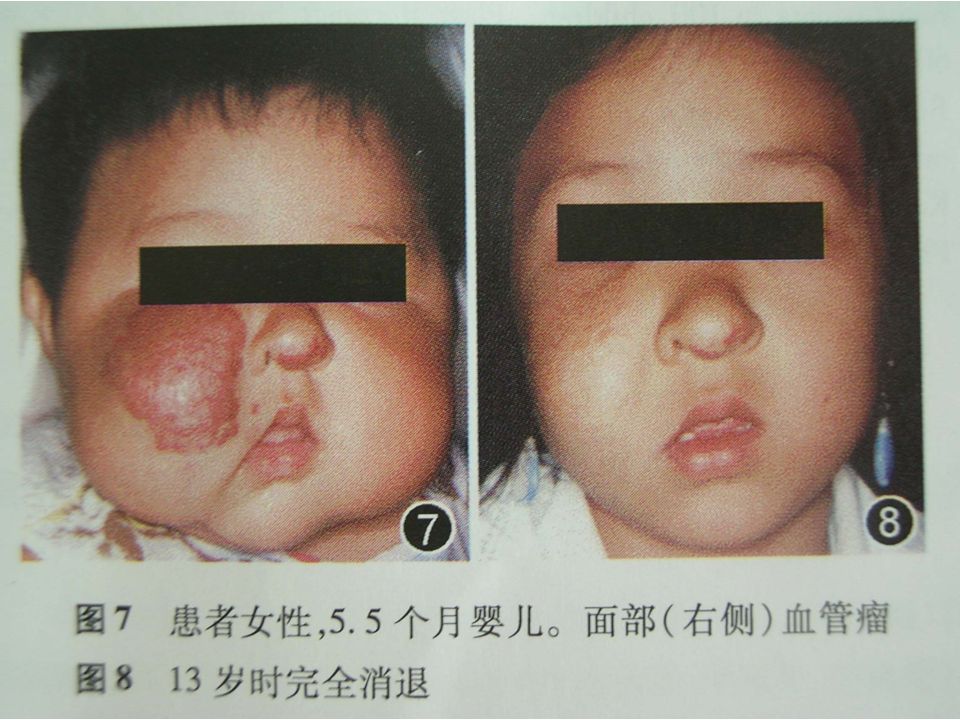
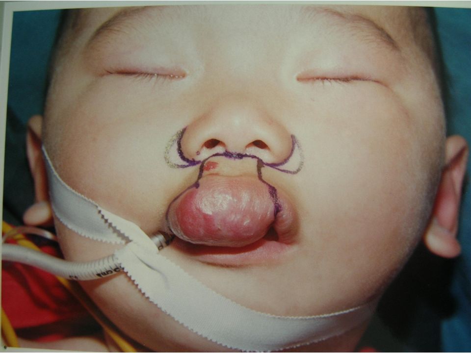
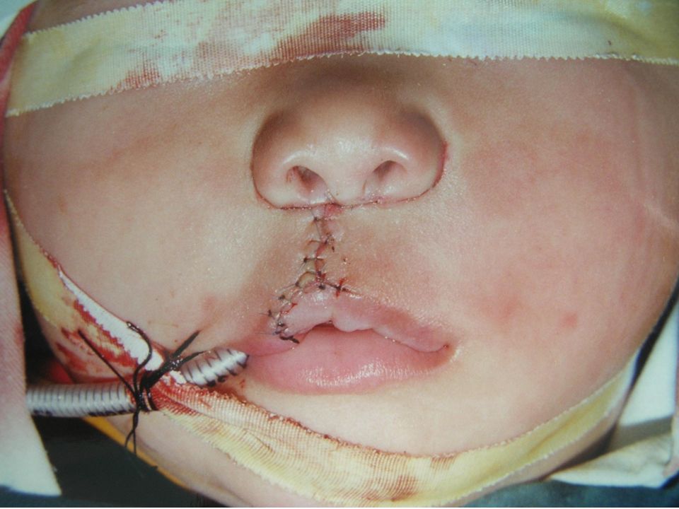
49
hemangioma 血管瘤 1) authentic tumor 2) account for 60% of body hemangiomas 3) head vs.body? 1:9? 4) most disappear before 10 year-old 5) surgery or not?
 most disappear before 10 year-old 5) surgery or not .")
50
Clinical characteristics 1) newborn—strawberry like on skin 2) rapid growth—after 1 month 3) stillness or shrinking—after 1 year 4) disappear—50-60% within 5 year; 75% within 7 year; 90 % within 10 year. 5) end of disappear—10-12 year Question: shall we recommend surgery? Advantage ? Disadvantage?
 end of disappear—10-12 year Question: shall we recommend surgery. Advantage . Disadvantage .")
52
vascular malformation A) venular( 微静脉) malformation -- 50~200um, former grape wine splash (原葡萄酒色斑) Clinic characteristics: Press—disappear, distinguish from nevus (血管痣)
 venular( 微静脉) malformation -- 50~200um, former grape wine splash (原葡萄酒色斑) Clinic characteristics: Press—disappear, distinguish from nevus (血管痣)")
53
sponge-like grape wine splash

54
B ) venous malformation (静脉畸 形) Former cavernous (sponge-like 原海绵状 ) hemangioma (old textbook) clinic characteristics: 1) Compress test 2) Head down test 3) Puncture 4) Vein calculi (stone) Question: is angiography (血管造影) helpful?
 venous malformation (静脉畸 形) Former cavernous (sponge-like 原海绵状 ) hemangioma (old textbook) clinic characteristics: 1) Compress test 2) Head down test 3) Puncture 4) Vein calculi (stone) Question: is angiography (血管造影) helpful")
57
venous malf - before & after surgery

58
c)Arterio-venous malformation Or: congenital (先天性) arteriovenous malf. Former grape-vine-like hemangioma 原蔓状血 管瘤. Outstretched artery and vein connecting directly, omitting capillary net. Clinic characteristics : 1) earthworm like; 2) throbbing/pulsate 3) souffle ( 吹风音, caused by vortex )
 earthworm like; 2) throbbing/pulsate 3) souffle ( 吹风音, caused by vortex ).")
59
1) surgery– once or several times 2) intra cavity injection—for venous malf., with Bleomycin, maximum 6mg/once. 3) hormone / isotope—for infant within 1 year-old. 4) laser light—for superficial lesion. 5) embolism of blood vessel—glutin( 明胶) sponge/ metal spring, reopen in a few days. 6) trans-catheter( 导管) arterial embolization—under DSA, dangerous. Question: can ligation of external carotid artery cure arteriovenous malf. ? Treatment methods
 hormone / isotope—for infant within 1 year-old. 4) laser light—for superficial lesion. 5) embolism of blood vessel—glutin( 明胶) sponge/ metal spring, reopen in a few days. 6) trans-catheter( 导管) arterial embolization—under DSA, dangerous. Question: can ligation of external carotid artery cure arteriovenous malf. Treatment methods.")
60
(四)、神经源性肿瘤 (神经鞘瘤 neurolemmoma ) 源于神经鞘膜,又称雪旺瘤 Schwannoma 。 临床表现:卵圆 / 圆形,光滑(包膜完整), 质地坚韧,沿神经轴侧向可动(纵向困难), 大者可发生黏液变,可抽出血样液体,但不 凝固。颈上部与颈动脉体瘤鉴别。 治疗:手术。神经干上可将外膜剖开剥离。
、神经源性肿瘤 (神经鞘瘤 neurolemmoma ) 源于神经鞘膜,又称雪旺瘤 Schwannoma 。 临床表现:卵圆 / 圆形,光滑(包膜完整), 质地坚韧,沿神经轴侧向可动(纵向困难), 大者可发生黏液变,可抽出血样液体,但不 凝固。颈上部与颈动脉体瘤鉴别。 治疗:手术。神经干上可将外膜剖开剥离。")
61
神经纤维瘤 neurofibroma 源于神经鞘细胞及纤维母细胞。 临床表现:面颈部皮肤棕色斑块,隆起, 扪诊质地柔软,可及多发性瘤结节,质 硬,感觉神经可有触痛,虽血运丰富但 不能压缩。多发:色斑 >1.5cm, >5~6 个时 ---- 神经纤维瘤病(遗传)。 治疗:手术。大出血准备。
。 治疗:手术。大出血准备。")
62
Before sugr. After sugr.

63
(五 ) 、骨源性肿瘤 osteogenetic tumor 骨化性纤维瘤(纤维组织多) 纤维骨瘤(骨组织多) 骨纤维异常增殖症(发育畸形)
 、骨源性肿瘤 osteogenetic tumor 骨化性纤维瘤(纤维组织多) 纤维骨瘤(骨组织多) 骨纤维异常增殖症(发育畸形)")
64
骨化性纤维瘤 ossifying fibroma 病理:大量束状和旋涡状纤维组织,部分不规 则的骨小梁和钙化团块。 临床表现:青年人,单发,下颌 > 上颌,女性 > 男 性,缓慢生长,质地硬, X 片界限清,密度减 低。 鉴别诊断:骨纤维异常增殖症 ---- 年龄更小,多发, 上颌 > 下颌, X 片界限不清,密度高低不等。 治疗:改善畸形 / 彻底切除。

65
(六)多形性腺瘤 唾液腺肿瘤中最常见的。又称混合瘤 ( mixed tumor). 临床表现:腮腺 > 颌下腺 > 舌下腺。结节 状肿块,缓慢生长。质地中等偏硬。可 恶变。 治疗:手术。
多形性腺瘤 唾液腺肿瘤中最常见的。又称混合瘤 ( mixed tumor). 临床表现:腮腺 > 颌下腺 > 舌下腺。结节 状肿块,缓慢生长。质地中等偏硬。可 恶变。 治疗:手术。")
66
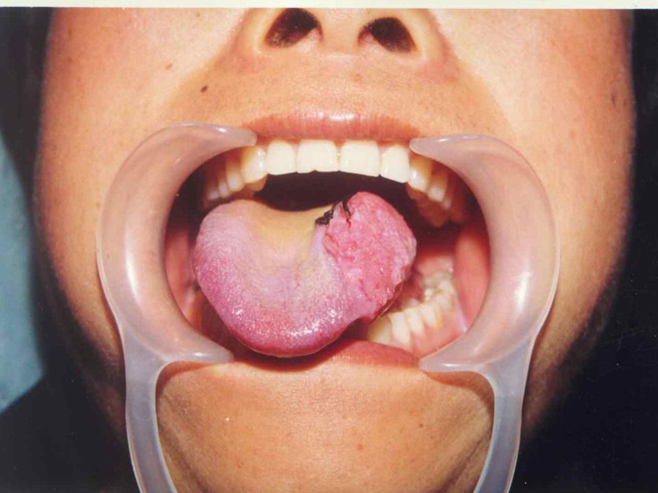
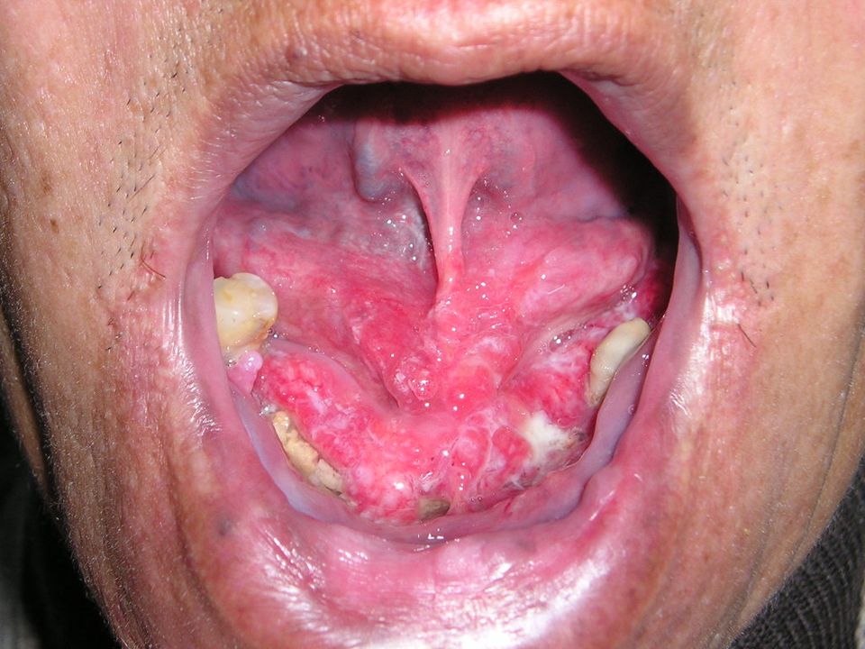
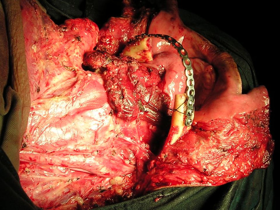
第四节 恶性肿瘤 一 、舌癌(约占口腔癌 30% ) (carcinoma of tongue) 病因:局部 - 刺激因素;全身 - 免疫监视 外因:物理,化学,生物 内因:免疫功能下降 临床表现:溃疡、外生、浸润三种类型。局部 通常有刺激因素。 早期:溃疡经久不愈。后 期:肿块长大各种表现。 治疗:综合治疗(手术、放疗、化疗、免疫、 中药)。手术:舌、颌、颈联合根治术。
 (carcinoma of tongue) 病因:局部 - 刺激因素;全身 - 免疫监视 外因:物理,化学,生物 内因:免疫功能下降 临床表现:溃疡、外生、浸润三种类型。局部 通常有刺激因素。 早期:溃疡经久不愈。后 期:肿块长大各种表现。 治疗:综合治疗(手术、放疗、化疗、免疫、 中药)。手术:舌、颌、颈联合根治术。")
68
二、牙龈癌 gingival (约 25% ):口腔卫 生差,农村多见。 三、颊癌 buccal (约 20% ):咬颊、白斑、 摩擦。 四、腭癌 palate ( 10% ):烟草性口炎、 百斑。 五、口底癌 oral floor :极易转移,预后 差。
:口腔卫 生差,农村多见。 三、颊癌 buccal (约 20% ):咬颊、白斑、 摩擦。 四、腭癌 palate ( 10% ):烟草性口炎、 百斑。 五、口底癌 oral floor :极易转移,预后 差。")
71
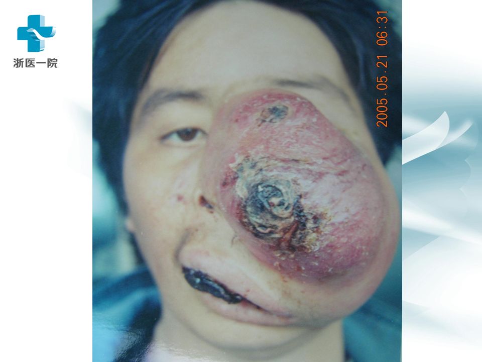
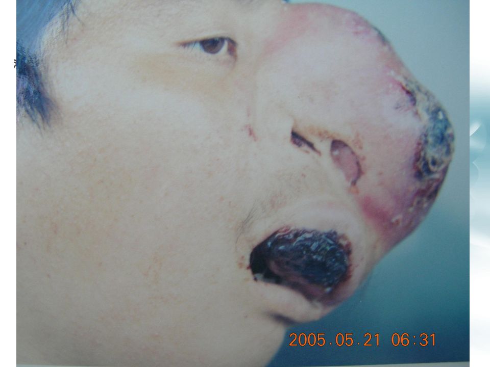
六、上颌窦癌 carcinoma of maxillary sinus 临床表现:早期无症状。后期因表现不同可能 首诊于不同科室。 1 )上颌窦底:牙、牙床 2 )内侧壁:鼻塞、鼻血 3 )上壁:突眼、复视 4 )前外壁:眶下神经受累、肿胀 5 )后外壁:张口受限 问题:上颌窦癌后期有那些临床表现?
上颌窦底:牙、牙床 2 )内侧壁:鼻塞、鼻血 3 )上壁:突眼、复视 4 )前外壁:眶下神经受累、肿胀 5 )后外壁:张口受限 问题:上颌窦癌后期有那些临床表现?")
74
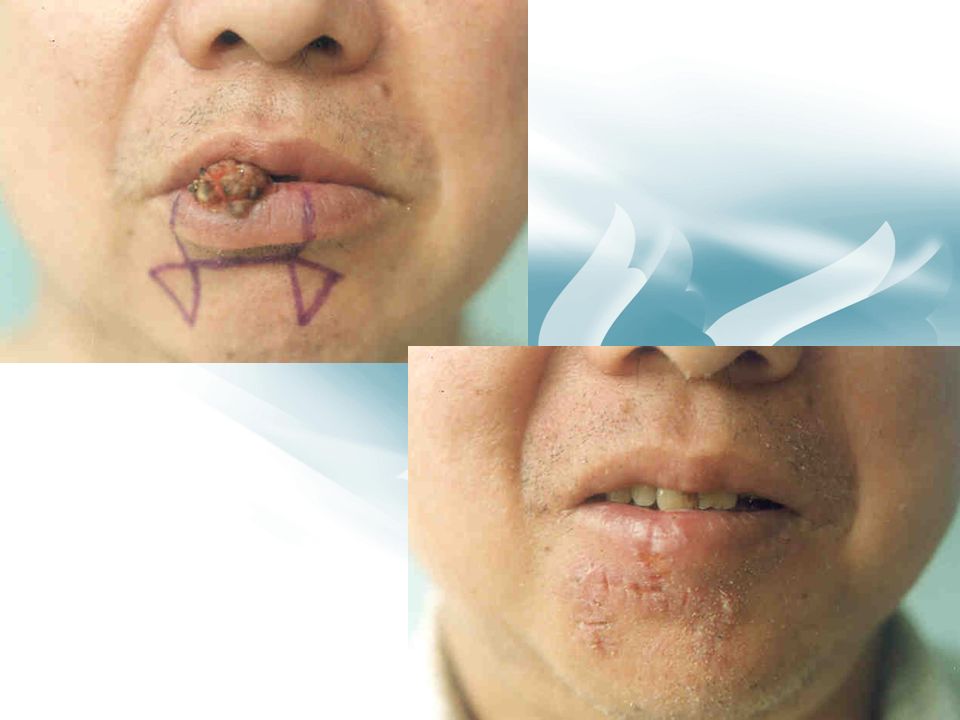
七、唇癌 carcinoma of lip 临床表现:唇红部火山口 crater 状溃疡 / 菜花状 cauliflower 肿块。 治疗:安全边界( 1 厘米)外切除。缺损 需要修复。预后好。
外切除。缺损 需要修复。预后好。")
76
八、口咽癌。九、颜面皮肤癌-基底细胞癌。 十、纤维肉瘤 fibrosarcoma :预后很差 十一、骨肉瘤 osteosarcoma :外伤史、颌骨破 坏、预后差 十二、恶性淋巴瘤 malignant lymphoma : HL : NHL ; 1 : 5 ;( NHL 预后差) 临床表现:多样性,颈部淋巴结肿大多见。 易误诊为炎症。靠免疫组化染色确诊。 治疗:化疗为主。
 临床表现:多样性,颈部淋巴结肿大多见。 易误诊为炎症。靠免疫组化染色确诊。 治疗:化疗为主。")
Similar presentations


. 2 基底细胞腺瘤( basal cell adenoma ) 多见于男性老年人,主要发生在腮腺.>")

 ? 口腔癌 -- 发生在口腔的恶性肿瘤 鳞状细胞癌 (Squamous cell carcinoma) 为 主 口腔:唇、舌、口腔底、颊黏膜、齿龈、 臼齿后区及颚部。 口腔癌在台湾之盛行率与致死率与日俱增, 为台湾十大癌症之第六名 ( 男性十大癌症发.>")


 第一节.>")
為然。因此,腫瘤是一種 有自主性( autonomous )的過渡發育或不正常新生.>")


 腹部肿块 Abdominal Mass.>")


