Download presentation
Presentation is loading. Please wait.

1
Osteoarticular and muscular system

2
骨关节系统最常规和 首选的检查方法

3
Bone and joint:Normal appearance of Radiology

6
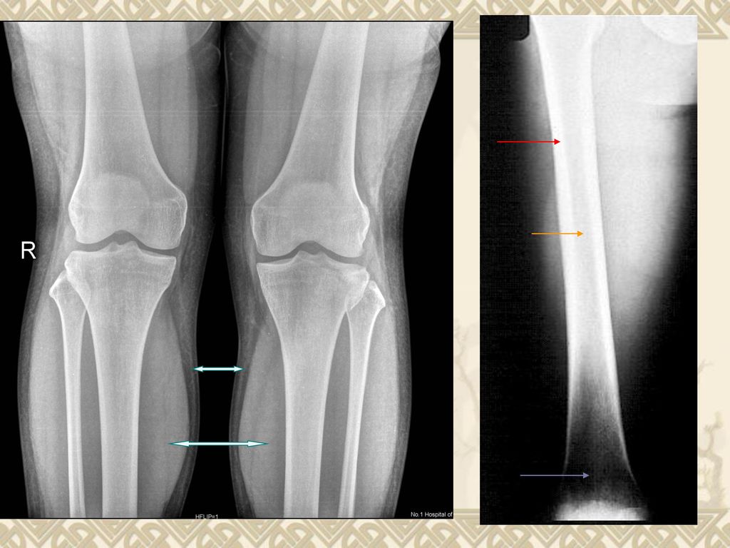
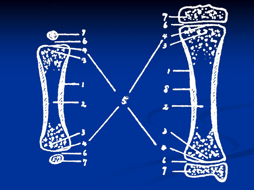
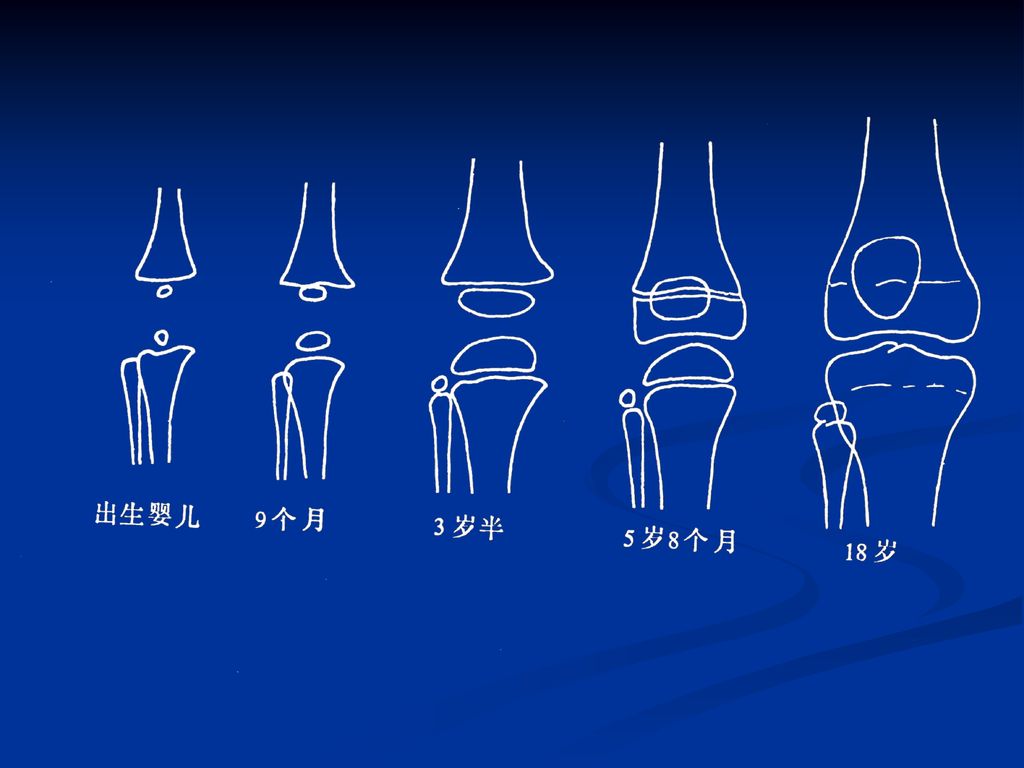
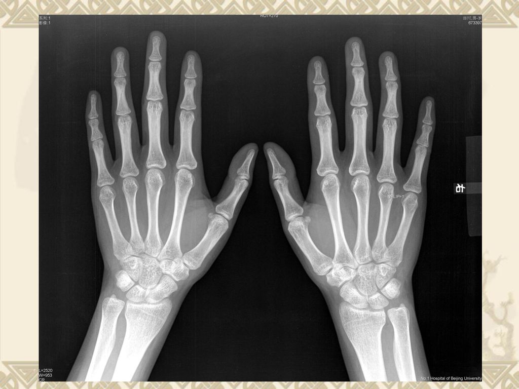
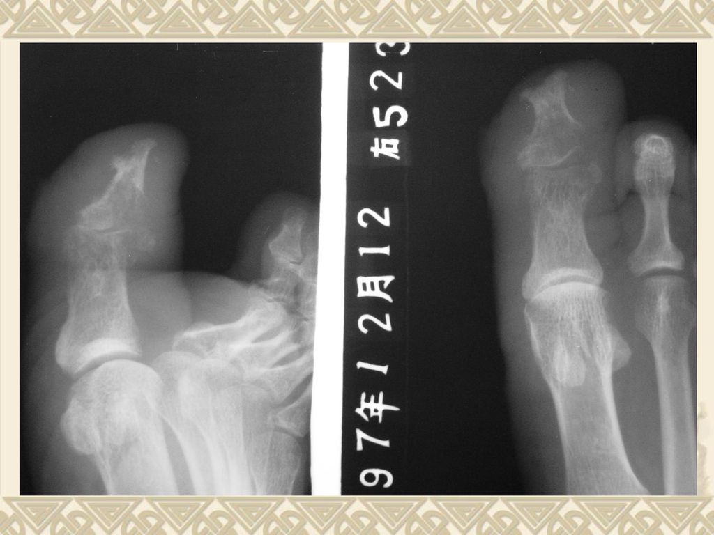
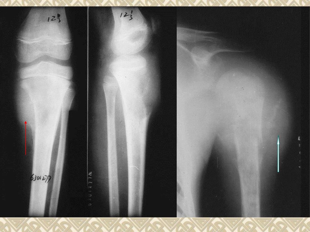
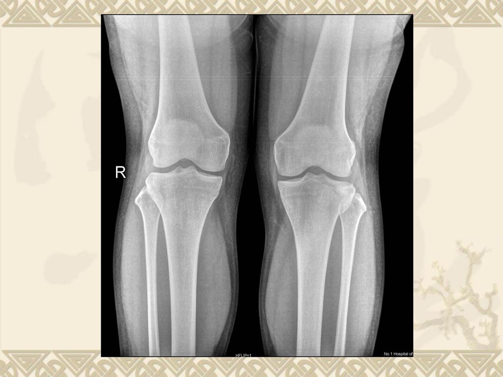
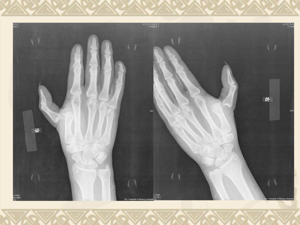
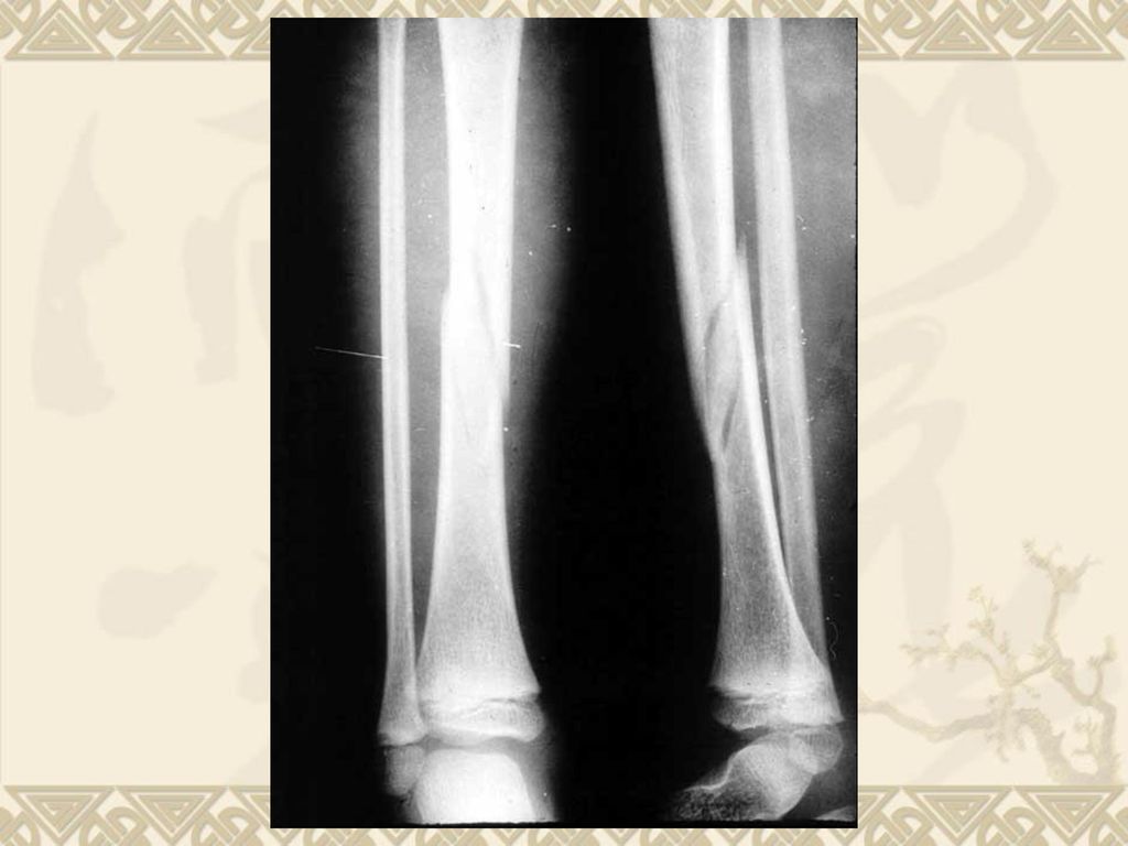
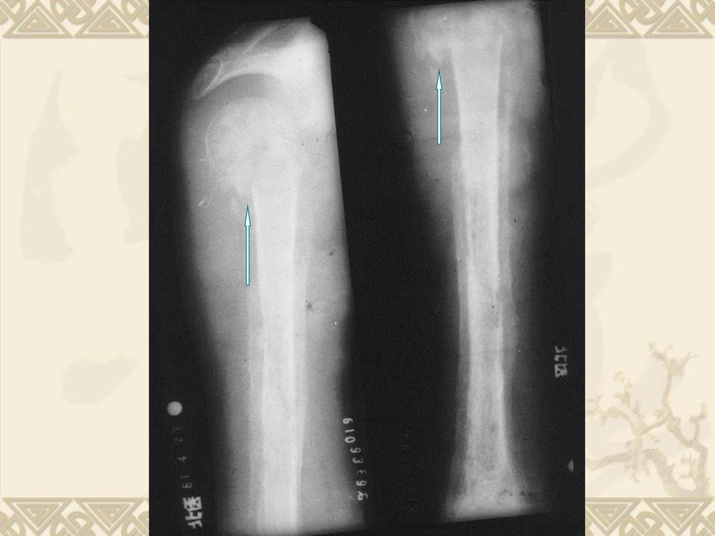
小儿长骨的特点:有未完全骨化的骺软骨

15
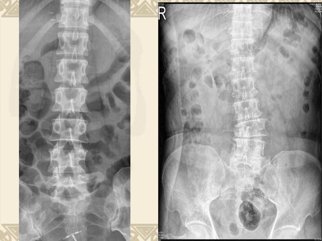
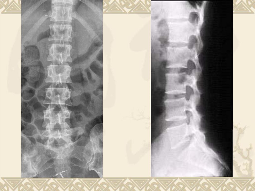
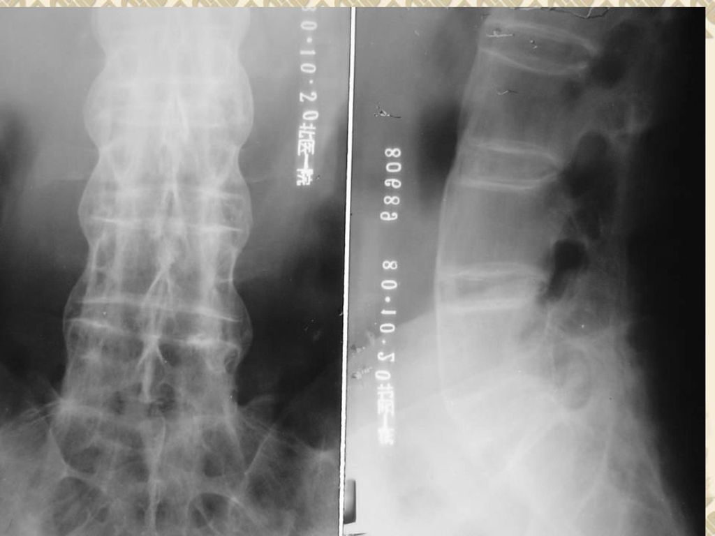
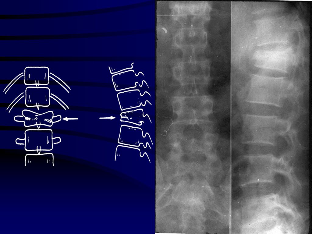
Spine 脊柱 生理曲度改变

16
The basic appearances of bone lesions

17
Osteoporosis Normal

18
Osteoporosis Normal

19
Normal Osteomalacia

20
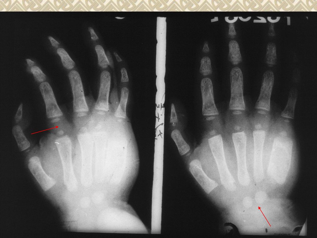
Destruction of bone

30
Hyperostosis and osteosclerosis
Normal

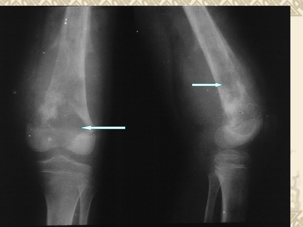
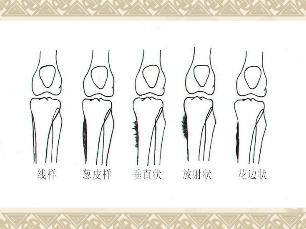
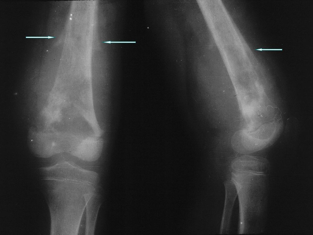
31
Periosteal proliferation 骨膜增生 Periosteal reaction 骨膜反应

35
Osteal and chondral calcification

37
Necrosis of bone

40
矿物质沉积

41
Deformation of bone

42
Surrounding soft tissue lesion

46
The basic appearances of joints lesions

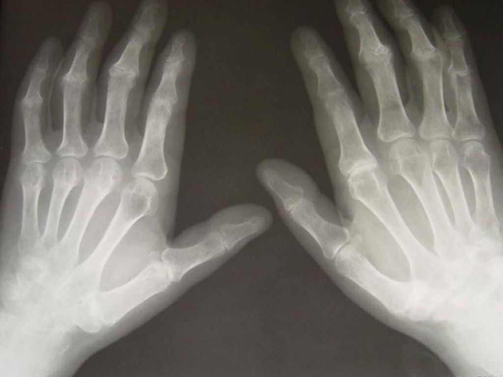
47
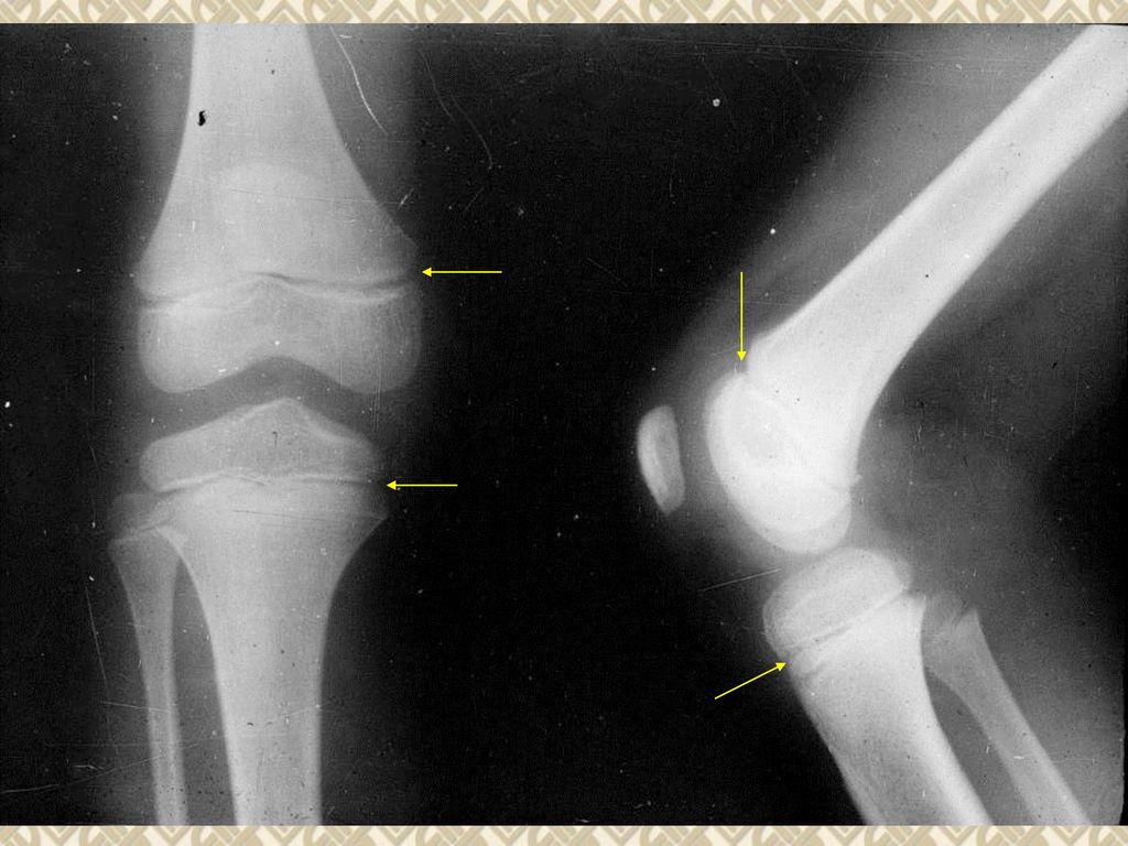
Degeneration of joint Destruction of joint Swelling of joint Ankylosis of joint Dislocation of joint

48
Degeneration of joint

49
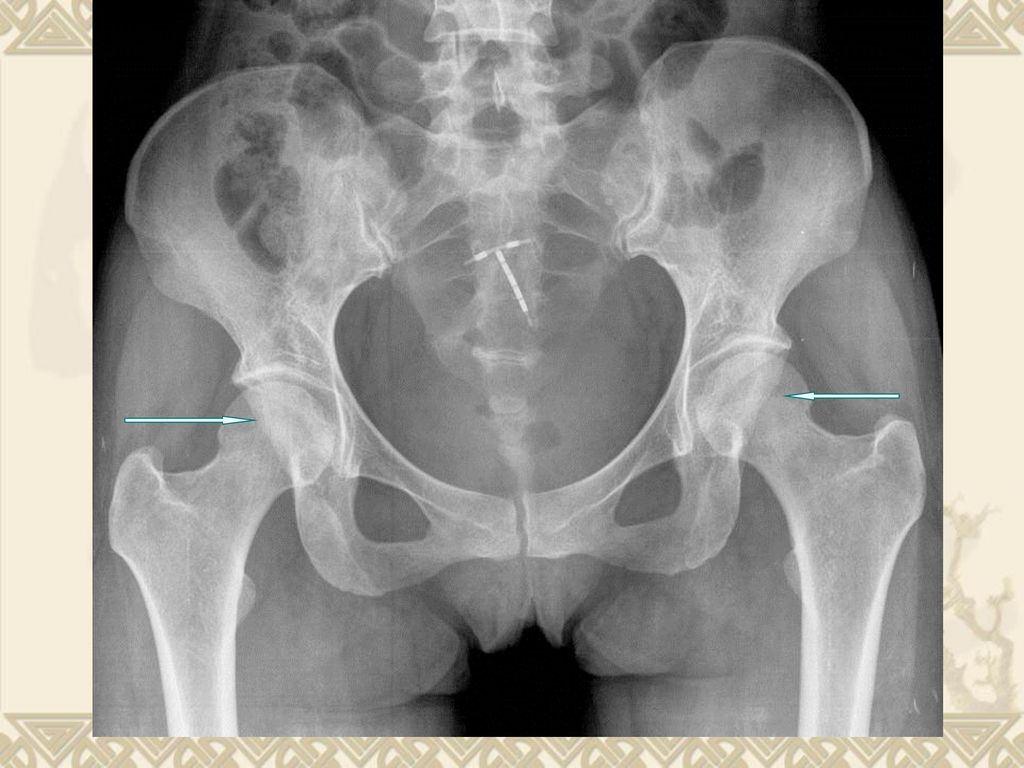
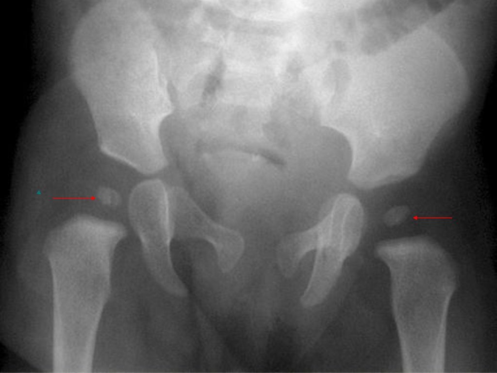
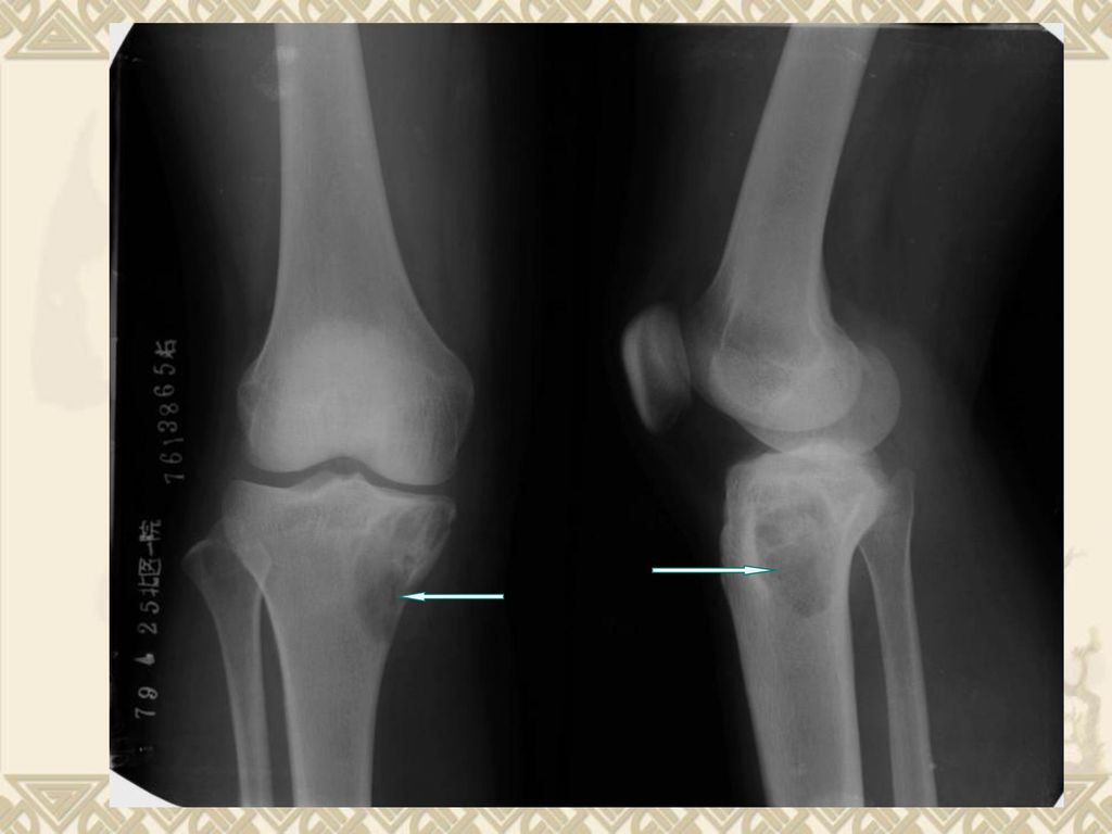
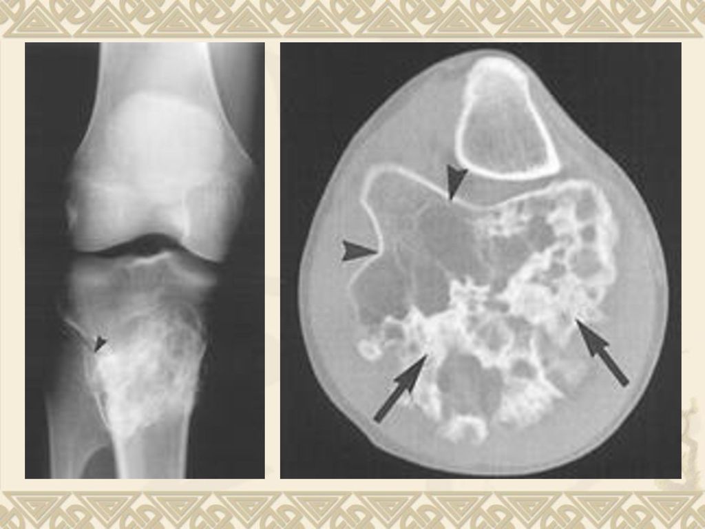
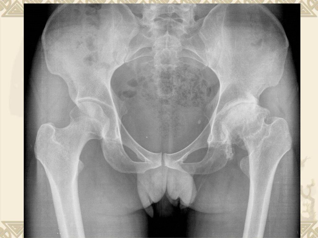
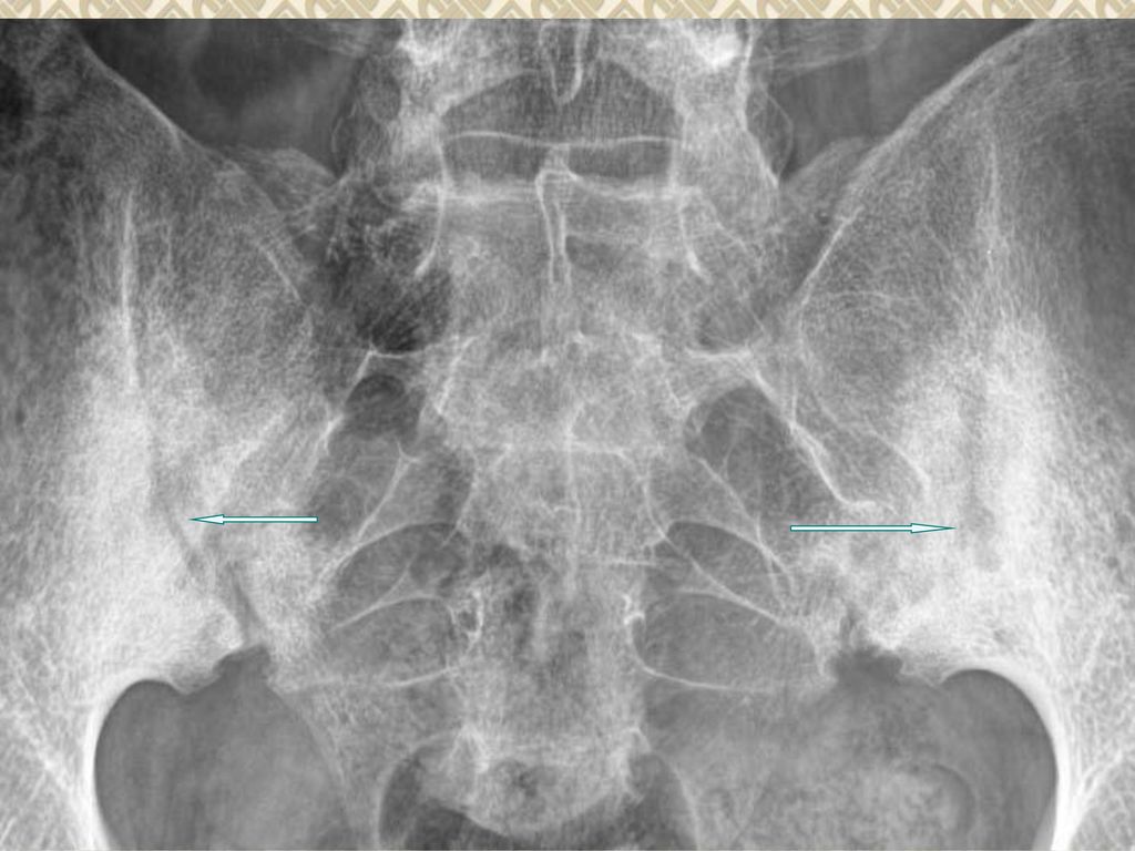
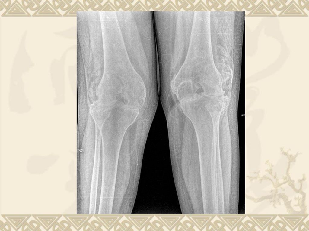
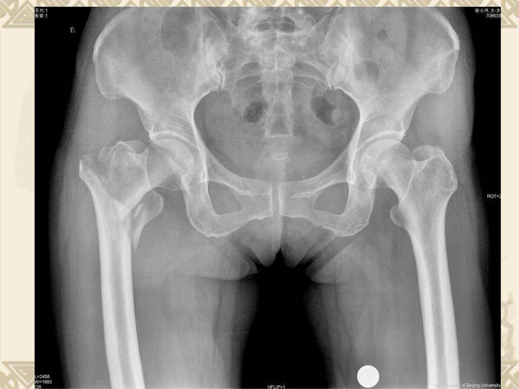
髋关节发育不良继发骨关节病

50
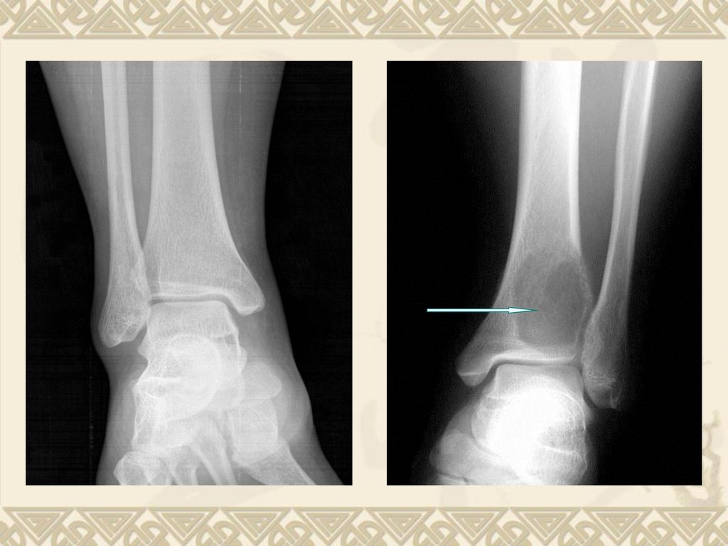
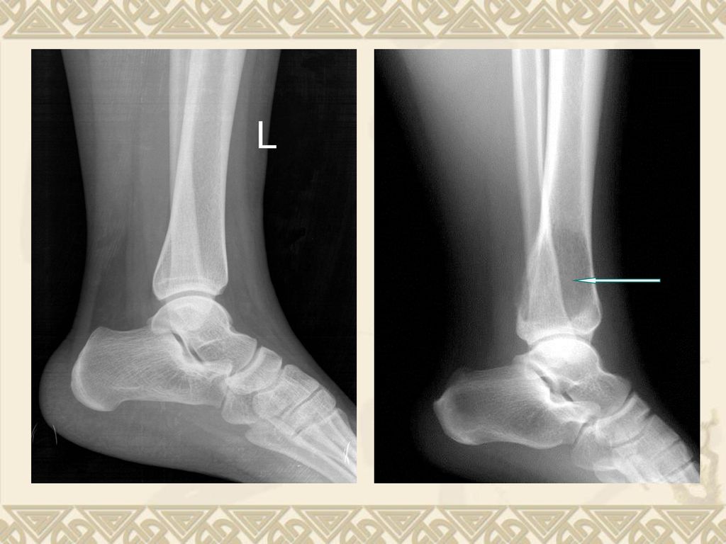
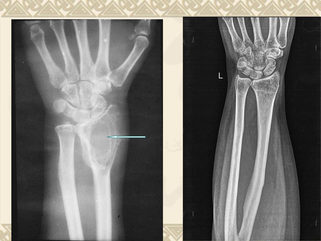
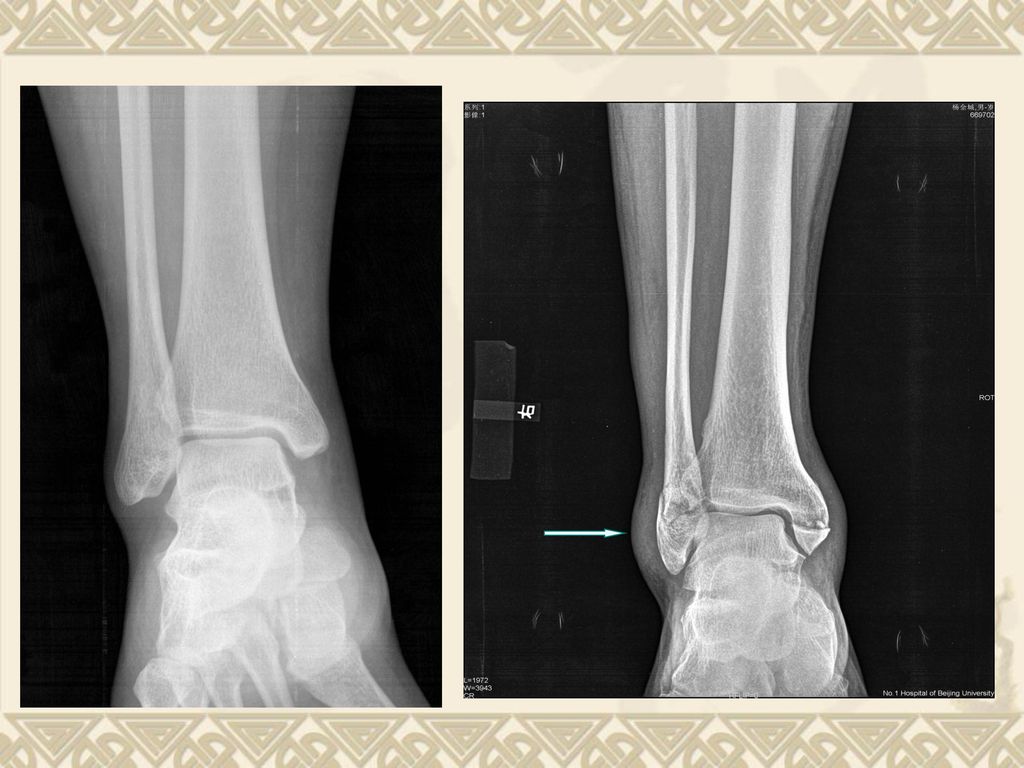
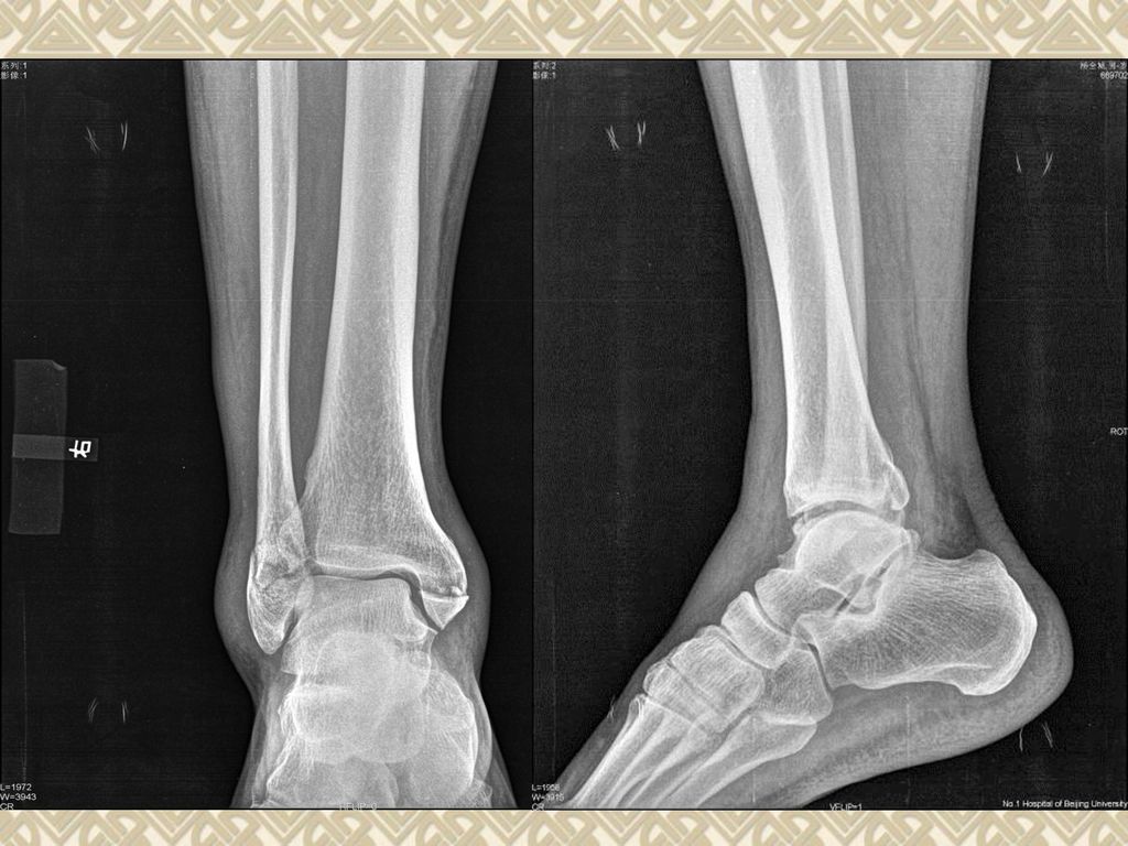
双踝退变

52
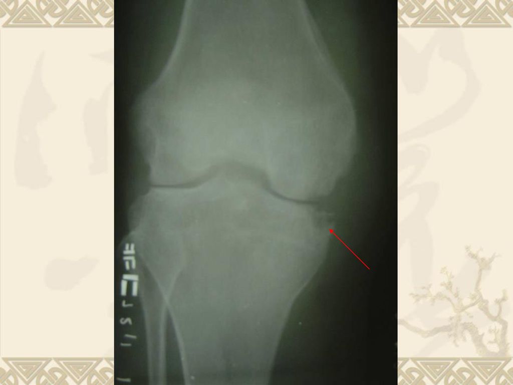
Destruction of joint

56
Swelling of joint

57
Swelling of joint Normal

60
Ankylosis of joint

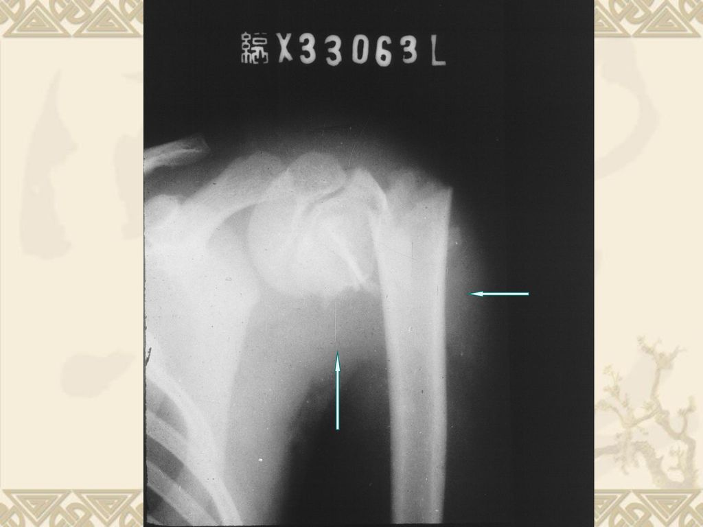
67
Dislocation of joint

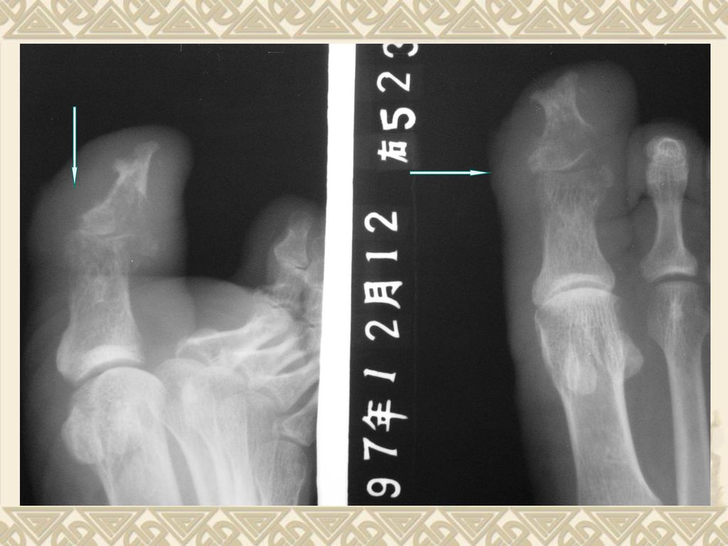
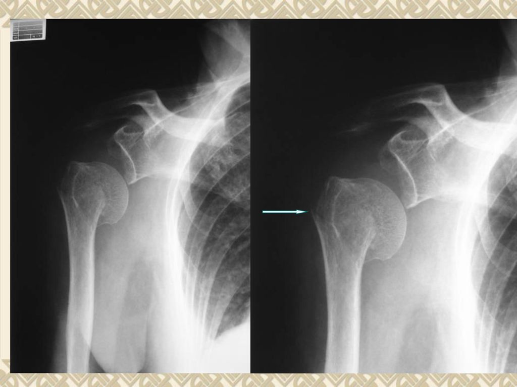
73
髌骨半脱位 髌骨轴位片

74
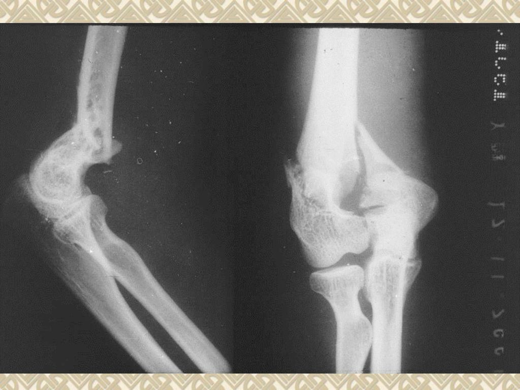
Fracture

75
对位------骨折复位断端对位不到1/2称对位不良; 对线-------骨折断端成角畸形称对线不良。

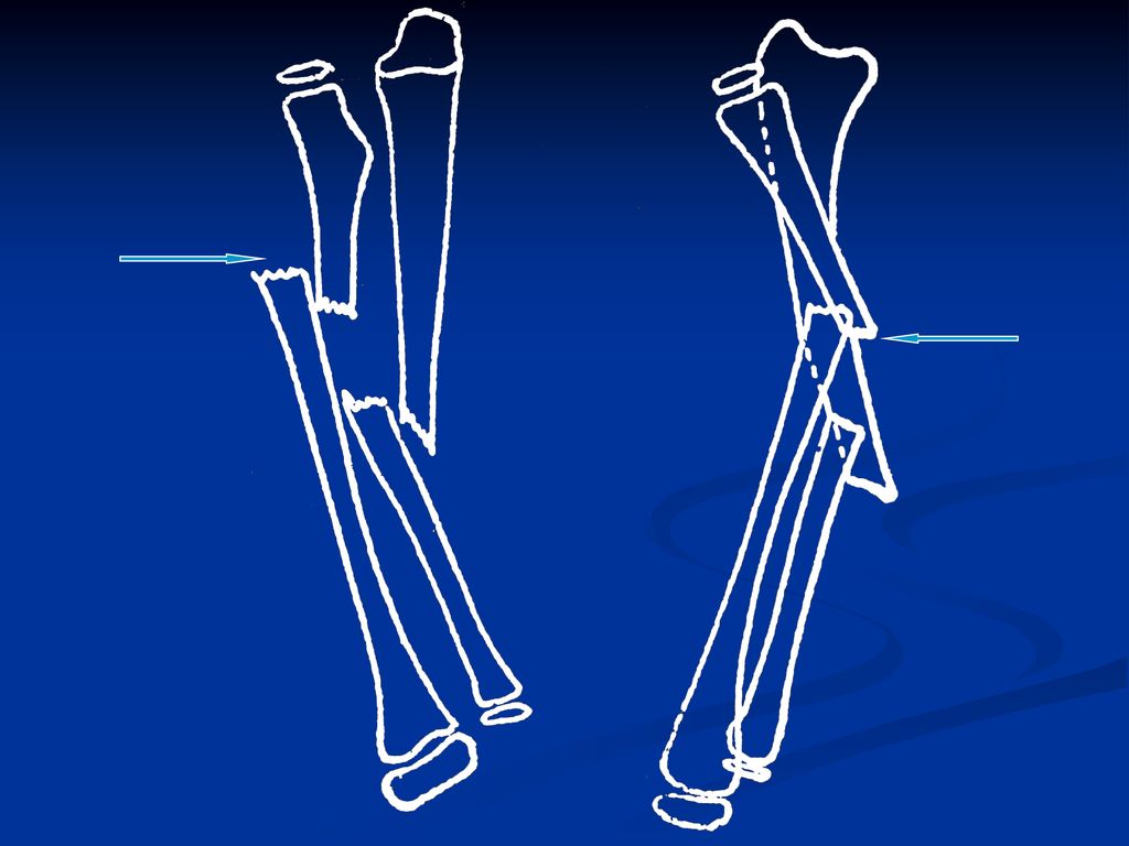
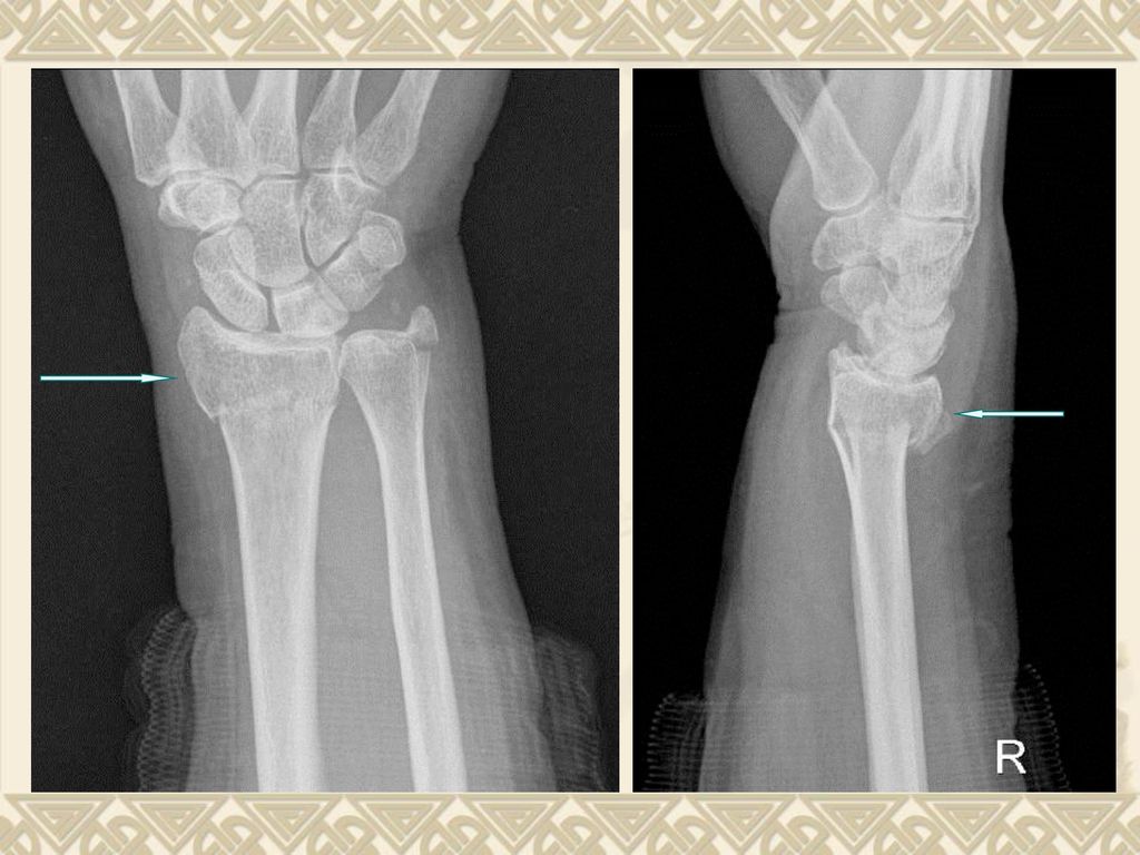
77
伸直型桡骨下端骨折(Colles骨折)
")
79
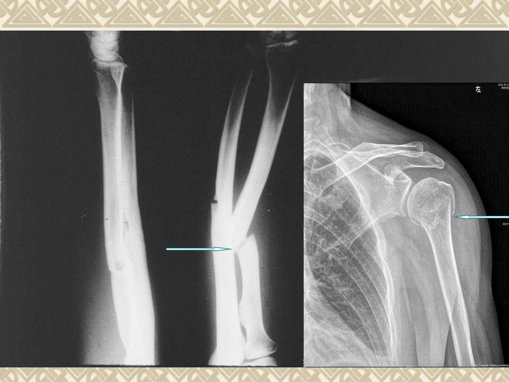
小儿青枝骨折(Green-stick fracture)
")
83
多发肋骨骨折

92
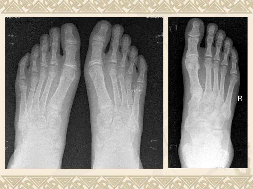
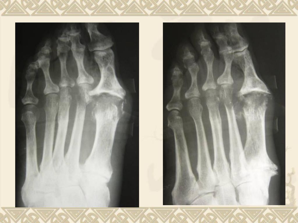
跖骨骨折

93
跟骨骨折

94
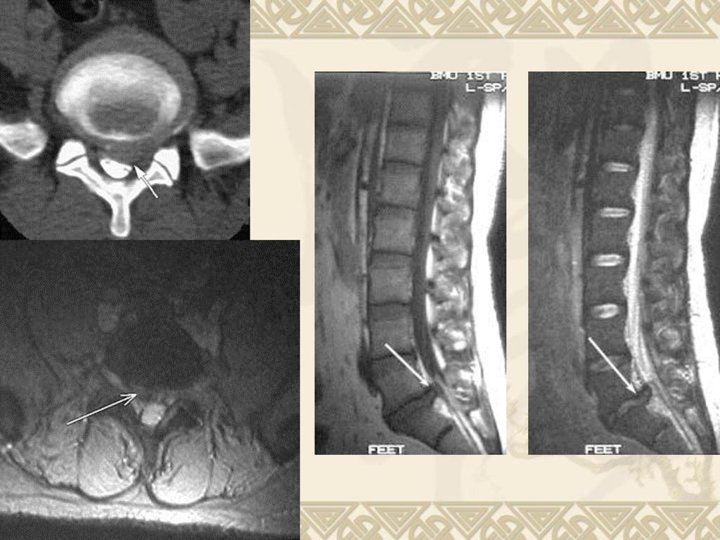
Abnormality of intervertebral disc

95
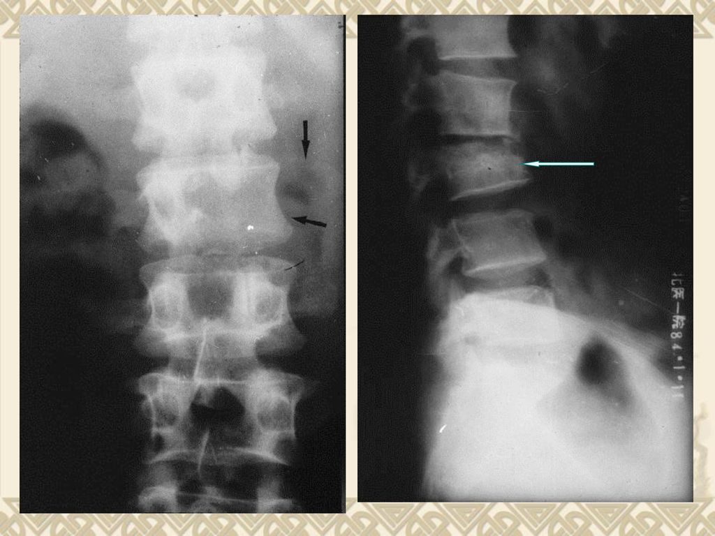
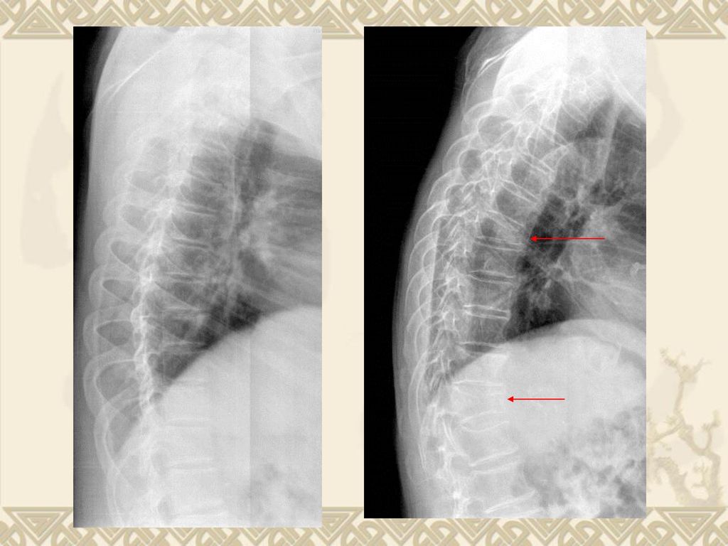
多发间盘病变、滑椎 椎间隙变窄

97
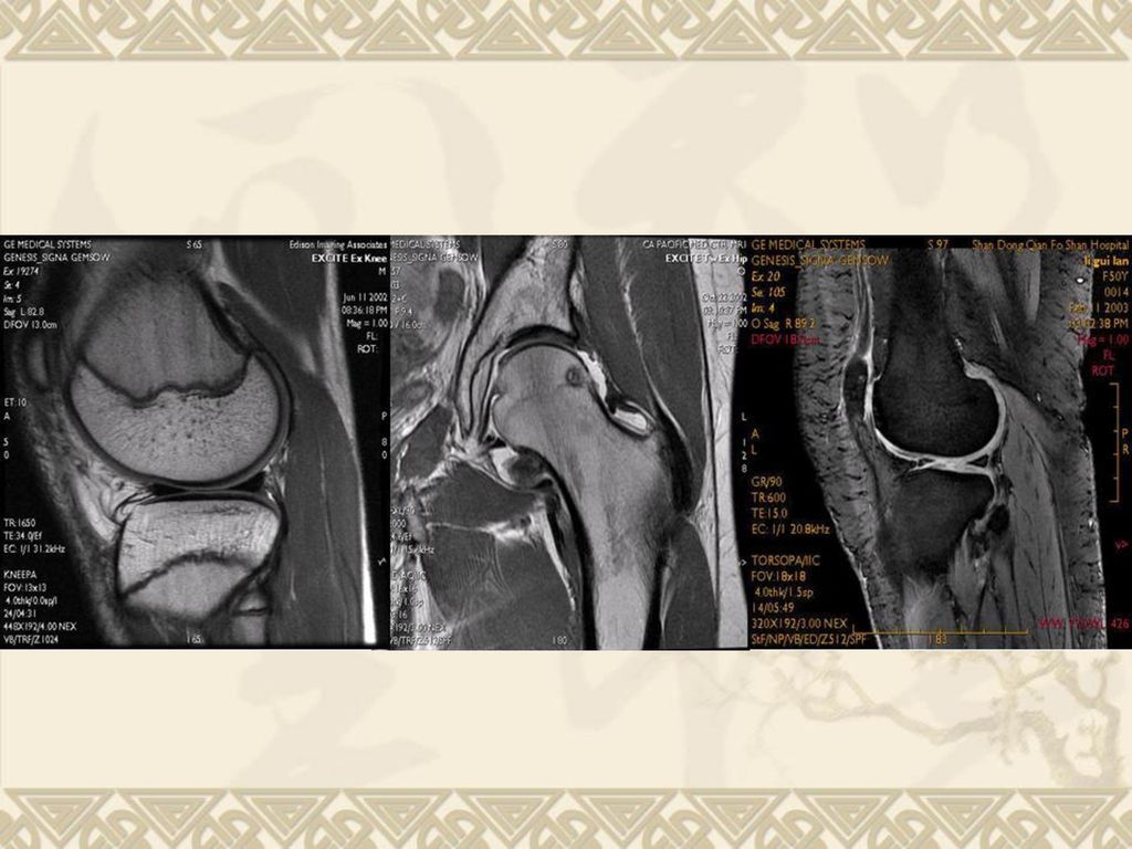
Trauma of joint dislocation of joint -- X线
injury of articular meniscus- injury of articular cartilage- injury of tendon and ligament- MRI MRI MRI

98
Joint

100
思考题 诊断骨关节系统疾病最常用、最常规和首选的检查方法: 判断题:
A. CT和MRI空间或密度分辨率高,软组织对比度好,断面成像避免重叠,可三维成像; B. CT和MRI对显示骨内小病灶、软组织、骨髓和关节内病变明显优于X光片; C. MRI能清晰显示脂肪、骨髓、韧带、肌腱、软组织、血管等正常结构和水肿、坏死、出血、肿瘤等病理改变,可显示关节内、骨髓腔和周围软组织结构的层次和形态。

101
思考题 目前显示关节内外结构和病理改变的最理想的方法: 显示脊柱解剖结构和病理改变以及了解病变与椎管内结构的关系的最佳影像学检查方法:
periosteal proliferation / periosteal reaction 可见于以下哪些情况: a. 炎症(骨髓炎); b. 恶性骨肿瘤(骨肉瘤); c. 外伤(骨折); d. 骨膜下出血;
; b. 恶性骨肿瘤(骨肉瘤); c. 外伤(骨折); d. 骨膜下出血;")
102
思考题 病理改变 基本病变 A. 正常骨质被病理组织所代替形成的 a. 骨质疏松 局部骨组织消失;
病理改变 基本病变 A. 正常骨质被病理组织所代替形成的 a. 骨质疏松 局部骨组织消失; B. 单位体积内正常钙化的骨组织减少, b. 骨质软化 即骨组织内有机成分和钙盐的含量 均减少,而二者的比例正常; C. 单位体积内骨组织的有机成分正常, c. 骨质破坏 而钙盐含量减少,即骨样组织钙化不足; D. 单位体积内的骨质含量增多; d. 骨质增生硬化 E. 血运障碍导致的骨组织局部代谢停止; e. 骨质坏死

103
思考题 关节间隙狭窄,软骨下骨质囊变、硬化,关节边缘骨赘形成是何种关节基本病变: 诊断骨关节创伤的首选检查方法:
判断肌腱、韧带断裂,复杂关节损伤和脊柱创伤常需进行的检查: 对位不良是指 ( ),对线不良是指( ); A. 横向和纵向移位( 分离和重叠 ); B. 断段成角;
,对线不良是指( ); A. 横向和纵向移位( 分离和重叠 ); B. 断段成角;")
104
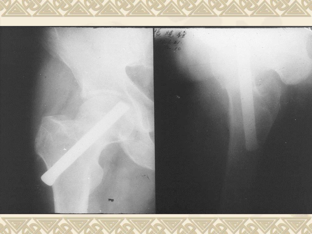
思考题 常见 fracture 特点 a. 股骨颈骨折; A. 桡骨远端2-3cm以内的横行或粉碎性fracture,
远侧断段向背侧或桡侧移位,断端向掌侧成 角畸形,可伴有尺骨茎突fracture; b. Colles’s fracture; B. 多见于老年人,常见于股骨头中部或基底 部,如影响股骨头血供,可导致愈合延迟或 股骨头缺血性坏死; c. 疲劳fracture C. 腓骨远端fracture、内踝基底或尖端撕脱 fracture、胫骨后缘fracture; d. 三踝fracture; D. 持续外力或长期积累性损伤所致的fracture, 常见于芭蕾舞演员、长跑运动员或战士;常 见部位为跖骨和胫骨;

105
思考题 儿童 fracture 的特殊类型 : a. green-stick fracture; b. 嵌插骨折; c. 压缩骨折;
d. 骺离骨折; fracture of spine X-ray平片可显示椎体形态的改变及骨碎片;要了解骨折片移位的程度、椎管的变形和狭窄、椎管内的碎骨片和血肿等,则需行何种检查;当脊柱fracture患者出现脊髓和神经根压迫症状,疑有脊髓压迫和脊髓挫裂伤时,需行何种检查;

106
思考题 椎间盘病变的常用影像学检查方法有: 临床上最常见的关节脱位: a. dislocation of shoulder ;
b. dislocation of elbow joint ; c. dislocation of hip joint; d. dislocation of knee ; e. dislocation of ankle ;

107
思考题 目前诊断injury of meniscus 和 injury of articular cartilage 首选的影像学检查方法:
判断injury of tendon and ligament 首选的影像学检查方法: 骨关节感染性疾病(化脓性骨髓炎、化脓性关节炎、骨与关节结核)最常规的影像学检查方法: 骨关节tumor和 tumor-like lesion 的常规和首选的影像学检查方法: 要了解恶性tumor 的侵犯范围和tumor与周围结构的关系,需要进行的进一步检查方法:
最常规的影像学检查方法: 骨关节tumor和 tumor-like lesion 的常规和首选的影像学检查方法: 要了解恶性tumor 的侵犯范围和tumor与周围结构的关系,需要进行的进一步检查方法:")
108
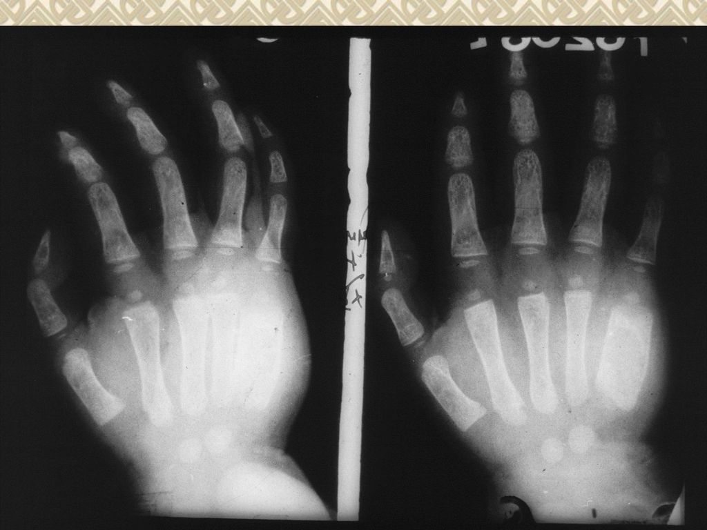
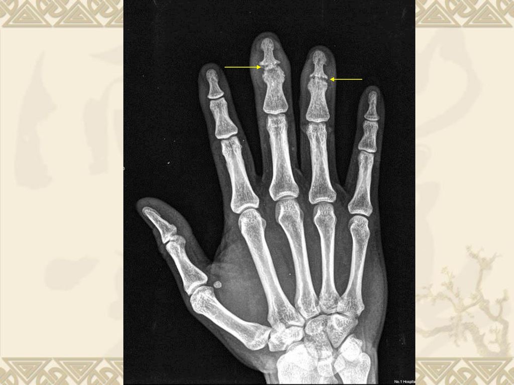
思考题 慢性骨关节病的典型表现是: a. 指间关节和掌指关节梭形肿胀,关节间隙变窄,关节面边缘侵蚀性骨破坏;晚期关节半脱位;
b. 小儿长管状骨干骺端内陷呈杯口状,干骺端边缘模糊呈毛刷状;下肢弯曲呈“O”形或“X”形腿; c. 成人全身骨质密度减低,椎体呈双凹变形,骨盆入口呈三角形变形; d. 关节间隙变窄,关节面硬化,边缘骨赘形成,晚期可见关节半脱位和关节内游离体;

Similar presentations


阻塞性肺不张 CT 表现为不张肺组织密度增高, 体积缩小,边缘清楚锐利,支气管阻 塞、中断,增强扫描明显强化,邻近 肺组织代偿性气肿,纵隔向健侧移位, 肺门移位。>")
