Download presentation
Presentation is loading. Please wait.

1
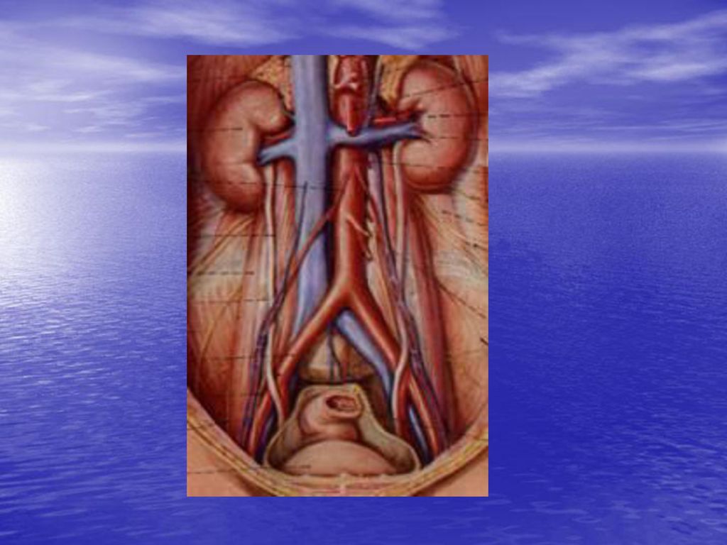
泌尿系统 urinary system 肾 输尿管 膀胱 尿道
泌尿系统 泌尿系统 urinary system 肾 输尿管 膀胱 尿道

2
Outline 重点 肾的位置和形态,以及肾门、肾蒂的概念,出入肾门的各结构的排列。掌握肾的构造。输尿管的起始和狭窄。掌握膀胱的形态、位置和毗邻。膀胱三角区境界和位置。 难点 肾被膜各层特点。膀胱分部和毗邻 掌握泌尿系统的组成及机能。 了解肾的被膜及固定结构,肾的细微结构。 了解输尿管的形态、位置、行程。掌握输尿管的起始和狭窄。 了解女性尿道的特点及开口位置。

3
肾kidney 是实质性器官,左右各一,位于腹后壁脊柱两侧,上端平第11-12胸椎体,下端平第3腰椎,后面贴腹后壁肌,前面被腹膜覆盖

4
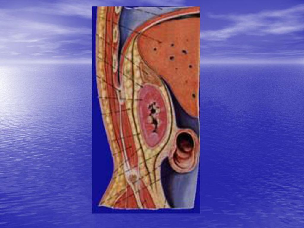
肾的位置及体表投影 正常成年人的肾位于腹膜后间隙内,脊柱的两旁,贴靠腹后壁的上部。肾的长轴向外下倾斜。右肾略低于左肾。左肾上端平第11胸椎下缘,下端平第2腰椎下缘。右肾上端平第12胸椎,下端平第3腰椎。第12肋斜过左肾后面的中部,右肾后面的上部。肾的位置存在个体差异,女性一般低于男性,儿童低于成人,新生儿肾位置更低,有时下端可达髂嵴附近。肾的正常位置靠多种因素来维持。肾被膜、肾血管、肾的邻接器官、腹内压及腹膜等。

5
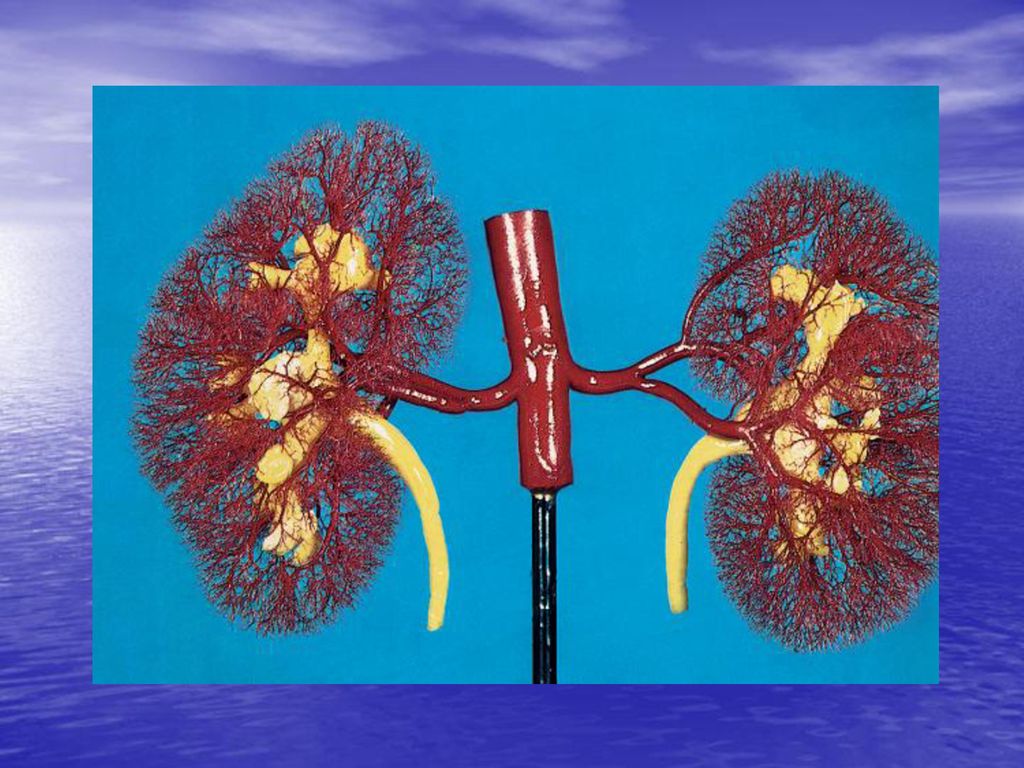
形态与结构 肾呈蚕豆形,分上下端,内外缘,前后面。内侧缘中部有血管、淋巴管、神经和肾盂出入称肾门。出入肾门的结构合称肾蒂。由肾门向肾内续于肾窦。窦内有肾动脉、肾静脉、肾小盏、肾大盏。肾小盏呈漏斗状,紧紧包绕着肾乳头,一个肾小盏包绕着1个或2个肾乳头,每2-3个小盏集合成肾大盏,大盏2-3个最后合并形成漏斗形的肾盂,出肾门后续于输尿管。

7
肾的冠状剖面上,可见肾实质分为皮质和髓质两个部分。肾髓质位于深部,色淡呈锥体形,叫肾锥体,锥体的尖端钝圆叫肾乳头。

8
Kidney Form: bean-shaped; 10 cm long, 5 cm wide & 4 cm thick; a superior and an inferior pole, a medial and lateral border, and an anterior and a posterior surface Location: T 11 or 12 to L3; right one slightly lower than left Relation: superior pole--adrenal; posterior--posterior abdominal wall; anterior--liver, duodenum & right colic flexure on right side, but spleen, tail of pancreas & left colic flexure on left side

9
Hilum: intermediate third of medial border where renal vessels, nerves, pelvis enter or leave kidney
Renal sinus : a space extending from hilum; which is occupied by renal pelvis, calices, vessels & nerves, & a variable amount of fat. Structures: 1)Renal cortex—glomeruli; between pyramids is called renal column 2)Renal medulla – 15 to 20 renal pyramids. Its apex--renal papilla 3)Minor calicos – cup-like; 7 to 8; surrounding 2 or 3 renal papillae. 4)Major calicos – formed by uniting of minor calicos; 2 or 3 5)Renal pelvis – formed uniting of major calicos; exiting kidney & turning downward to lead to ureter.
Renal cortex—glomeruli; between pyramids is called renal column. 2)Renal medulla – 15 to 20 renal pyramids. Its apex--renal papilla. 3)Minor calicos – cup-like; 7 to 8; surrounding 2 or 3 renal papillae. 4)Major calicos – formed by uniting of minor calicos; 2 or 3. 5)Renal pelvis – formed uniting of major calicos; exiting kidney & turning downward to lead to ureter.")
10
Kidneys Position: 1) Retroperitoneal space 2) T11-L2 (left),
T12-L3(right)
11
Adjacent organs

13
Structure Renal hilum--renal pedicle Renal sinus
1) 2 surfaces, 2 margins, 2 poles 2). Renal hilum-- (1). At the media margin (2). A vertical cleft (3). Renal a., v., renal pelvis, N., L. and connective tissue. entering and leaving the Renal hilum--- renal pedicle (4). Relationship of these structures---V., A.,Pelvis anteroposterorly 3). Renal sinus Renal hilum--renal pedicle Renal sinus
 2 surfaces, 2 margins, 2 poles. 2). Renal hilum-- (1). At the media margin. (2). A vertical cleft. (3). Renal a., v., renal pelvis, N., L. and connective tissue. entering and leaving the Renal hilum--- renal pedicle. (4). Relationship of these structures---V., A.,Pelvis anteroposterorly. 3). Renal sinus. Renal hilum--renal pedicle. Renal sinus.")
14
Minor calix 肾小盏 Major calix Renal pelvis Renal a. and v. N. Fat

15
Coronary section Renal cortex (肾皮质) Renal column Renal medulla(肾髓质) Renal pyramids(15-20) Renal papilla
 Renal pyramids(15-20) Renal papilla.")
16
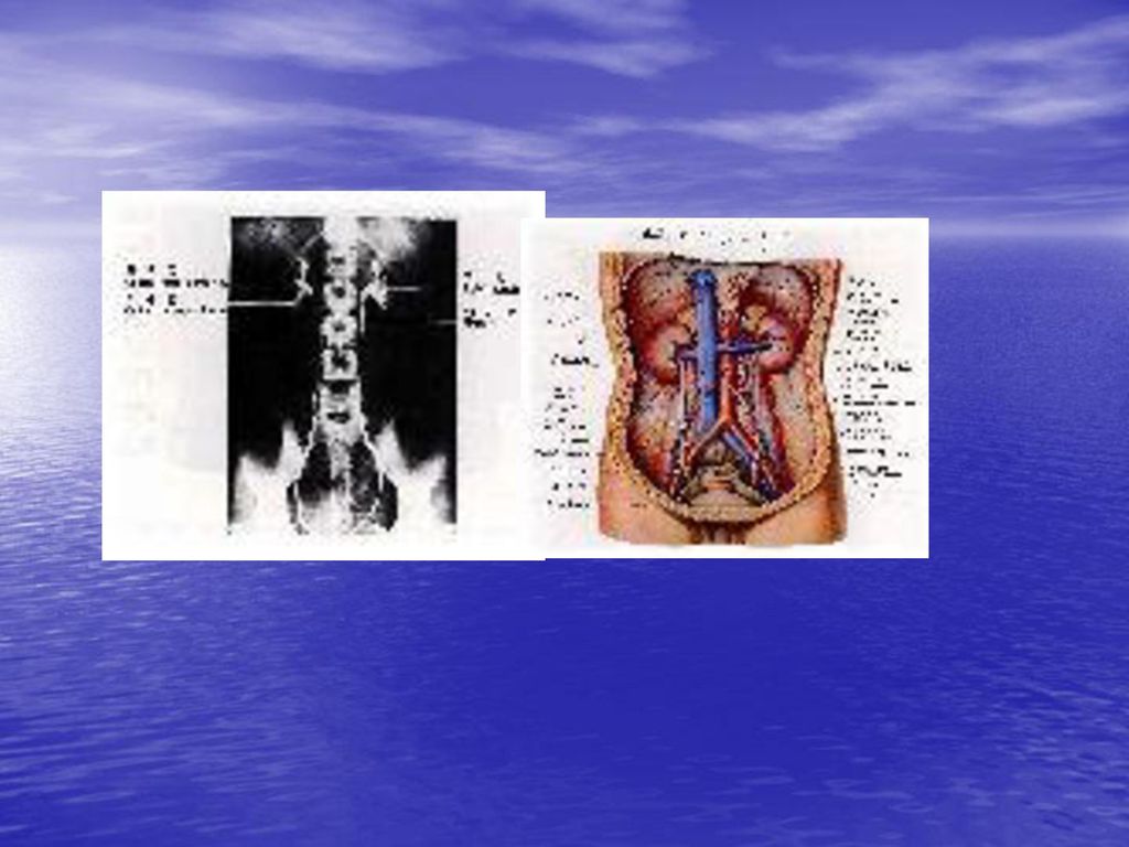
Renal vessels and renal segments

18
Variations of renal artery

19
Variations of renal veins

20
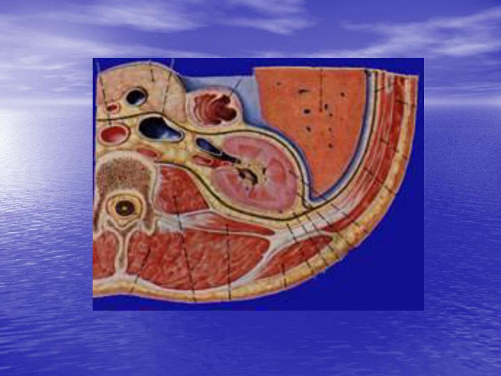
肾表有三层被膜,由外向内分别为,肾筋膜、脂肪囊、纤维囊

23
1. 肾筋膜:穿过脂肪囊与肾纤维膜相连,具有保护固定肾脏的作用
前层:肾前筋膜 后层:肾后筋膜,肾后筋膜经肾后方与腰大肌和腰方肌的筋膜密接,肾后筋膜向下与骼嵴与髂筋膜愈着 外侧:肾的外缘互相融合 内侧:肾前筋膜越过肾脏的前方与对侧相连续 肾上方:二层于肾上腺的上端相互结合,与膈下筋膜相连接,肾下方肾前筋膜消失于腹膜下组织中,肾筋膜下端开放,形成肾下垂(游走肾)
")
24
2. 肾脂肪囊:称肾床,为脂肪组织层 3. 肾纤维膜:为肾固有膜,保护肾实质的作用

25
RENAL FASCIA AND FAT Fibrous capsule connects kidney loosely
Perirenal fat is continuous at hilum of kidney with fat in renal sinus Renal fascia is fibrous tissue surrounding kidney. It sends bundles of collagen through fat, which, along with the renal vessels and ureter, hold kidney in position. It ascends to envelop suprarenal glands & fuses as infraphrenic fascia. Inferior to kidney it is replaced by loose connective tissue. Lateral to kidney it fuses & continues with transverse fascia. Medially its anterior layer crosses great vessels to continue with opposite one; posterior layer is fused with fascia of vertebral column.

26
Covers of the kidney Fibrous capsule Perirenal fat Renal fascia

27
出入肾门所有的结构,即出入肾门的输尿管,肾A,肾V和神经、淋巴管 各结构的排列关系: 由前向后:依次为肾V、肾A、输尿管
肾蒂的概念: 出入肾门所有的结构,即出入肾门的输尿管,肾A,肾V和神经、淋巴管 各结构的排列关系: 由前向后:依次为肾V、肾A、输尿管 由上向下:依次为肾A、肾V和输尿管

28
输尿管ureter: 长约30厘米,自肾盂起始后,首先沿腹后壁下行,再沿盆腔侧壁至盆底向内下斜穿膀胱壁,开口于膀胱。输尿管分三段,即腹段、盆、膀胱壁内段

31
输尿管有三个狭窄: 1.起始部(与肾盂交接处) 2.与髂血管交叉处(跨髂外A处) 3.膀胱壁内段
 2.与髂血管交叉处(跨髂外A处) 3.膀胱壁内段")
32
输尿管有三个交叉: 1.与生殖腺血管交叉 2.与髂外血管交叉 3.与子宫动脉(输精管)交叉
交叉")
33
Ureter A pair of 25 cm long muscular ducts with narrow lumina carrying urine from the kidneys to urinary bladder Abdominal part is retroperitoneal through-out its course & runs inferomedially along transverse processes of lumbar vertebrae & crosses external iliac artery Pelvic part runs along lateral wall of pelvis to enter urinary bladder.

34
Ureter 1. Muscular ducts 2. 3 parts—abdominal , pelvic , and intramural parts 3. 3 narrow constrictions 1) at the junction of the ureter and renal pelvis 2) crossing sup. aperture of lesser pelvis 3) intramural part
 at the junction of the ureter and renal pelvis. 2) crossing sup. aperture of lesser pelvis. 3) intramural part.")
35
膀胱urinary bladder 上连输尿管,下接尿道。位于小骨盆腔内,前为耻骨联合,后方在男性有精囊腺、输精管和直肠,在女性有子宫和阴道
膀胱空虚时呈锥形,分膀胱尖、膀胱底、膀胱体、膀胱颈。在膀胱底内面有膀胱三角。三角的三顶角分别是尿道内口和左右输尿管开口。在左右输尿管口之间有输尿管间襞

36
膀胱空虚时,其内粘膜面呈现许多皱襞,唯其底部有一三角形的平滑区,称膀胱三角,两侧角即左、右输管口,两口之间有呈横向隆起的输尿管间襞,三角的前下角为尿道内口,膀胱三角是结核与结石等的好发部位。

37
1、膀胱的位置,毗邻及机能状态 膀胱的位置,随年龄及盈虚状态而不同,空虚时呈锥体状,位于盆腔前部,可分尖、体、底、颈四部,但各部间无明显分界。充盈时可升至耻骨联合上缘以上,此时腹膜返折处亦随之上移,膀胱前外侧壁则直接邻贴腹前壁。临床上常利用这种解剖关系,在耻骨联合上缘上进行膀胱穿刺或做手术切口,可不伤及腹膜。儿童的膀胱位置较高,位于腹腔内,到六岁左右逐渐降至盆腔。

38
空虚的膀胱,前方与耻骨联合相邻,其间为耻骨后隙,膀胱下外侧面邻肛提肌,闭孔内肌及其筋膜,其间充满疏松CT等,称膀胱旁组织,内有输尿管盆部,男性还有输精管壶腹穿行。膀胱后方借直肠膀胱隔与精囊、输精管壶腹及其后方的直肠相邻,女性则借膀胱阴道隔与子宫颈及阴道前壁相邻,膀胱上面覆盖腹膜,并与肠袢相邻,女性还与子宫相邻。膀胱的后下部即膀胱颈,下接尿道。男性邻贴前列腺,女性与尿生殖膈相邻。

40
尿道urethra 是排尿管道的最后一段,由膀胱下口(尿道内口)开始,末端直接开口于体表。 男、女尿道有很大不同。
男性尿道male urethra细长曲 女性尿道female urethra短阔直

41
男性尿道特点: 功能:具有排精和排尿的功能 特点: 1)3处狭窄 尿道内部;尿道膜部;尿道外部 2)3扩大:前列腺部;尿道球部;尿道舟状窝 3)两个弯曲:耻骨下曲:较为固定;耻骨前曲:阴茎上提时此弯曲可消失
3扩大:前列腺部;尿道球部;尿道舟状窝. 3)两个弯曲:耻骨下曲:较为固定;耻骨前曲:阴茎上提时此弯曲可消失.")
Similar presentations


:動物體藉各器官、系統排 除體內代謝廢物的過程 新陳代謝廢物以含氮廢物(尿素、尿酸)為主, 此外,尚有二氧化碳、多餘水分及熱能等.>")


 基本内容:肾和排尿管道的一般结构、肾单位的组织结构与尿液生成的关系。>")


 计算机断层摄影.>")